
Human Respiratory System
1. Introduction & Respiratory Organs Across Animals
A. Basic Definitions
- Breathing (Pulmonary Ventilation): Physical process of gaseous exchange — Inhalation ($O_2$-rich air in) and Exhalation ($CO_2$-rich air out).
- Respiration: Biochemical process involving the oxidation of food (glucose) inside cells to release energy (ATP).
B. Respiratory Organs in Animal Kingdom
| Animal Group | Respiratory Organ / Mechanism |
|---|---|
| Lower Invertebrates (Sponges, Coelenterates, Flatworms) | Simple diffusion over entire body surface (No specialized structure). |
| Annelids (Earthworm) | Moist, vascular, thin cuticle skin (Cutaneous respiration). |
| Insects (Cockroach) | Tracheal tubes & Spiracles (Tracheal system). |
| Aquatic Arthropods & Molluscs / Fishes | Vascularized Gills (Branchial respiration). |
| Amphibians (Frog) | Gills (larva), Moist Skin (Cutaneous), Buccal Cavity, and Lungs (Pulmonary). |
| Reptiles, Birds, Mammals | Vascularized Lungs (Pulmonary respiration). |
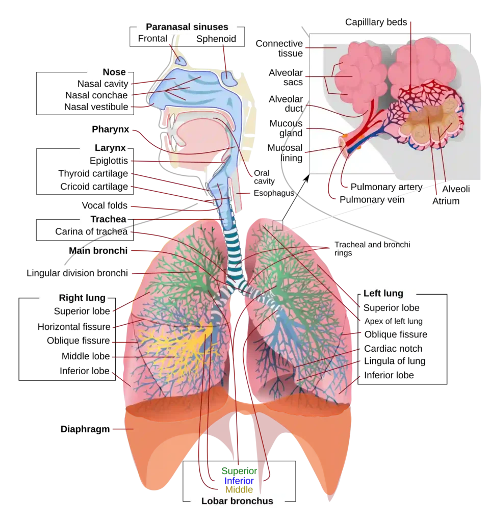
C. Human Respiratory System Pathway
- External Nares (Nostrils): Paired openings at the base of the nose.
- Nasal Cavity: Divided by nasal septum. Contains:• Vestibule: Hairs and oil glands to trap dust particles.
• Respiratory Region: 3 nasal conchae (bony projections) for conditioning and warming air.
• Olfactory Region: Lined by Schneiderian membrane for sense of smell.
- Pharynx: Common passage for food and air. Divided into Nasopharynx, Oropharynx, and Laryngopharynx.
- Larynx (Voice Box): Cartilaginous structure made of 9 cartilages:• Unpaired (3): Epiglottis (elastic), Thyroid (largest, Adam’s apple), Crico-thyroid/Cricoid (signet ring-shaped).
• Paired (3): Arytenoid, Corniculate, Cuneiform.
• Vocal Cords: 1 pair false (upper), 1 pair true (lower). Post-puberty testosterone makes male vocal cords thick and long $\to$ Low pitch voice.
2. Tracheobronchial Tree, Lungs & Breathing Mechanism
A. Trachea & Bronchial Tree Division
- Trachea (Windpipe): Straight tube lined by pseudostratified ciliated columnar epithelium. Supported by 16–20 incomplete C-shaped hyaline cartilaginous rings to prevent collapse during low pressure.
- Divides at the level of 5th Thoracic Vertebra ($T_5$) into Primary ($1^\circ$) Right and Left Bronchi.
- Division Branching:$$\text{Trachea} \to 1^\circ \text{Bronchi} \to 2^\circ \text{Bronchi} \to 3^\circ \text{Bronchi} \to \text{Terminal Bronchiole} \to \text{Respiratory Bronchiole} \to \text{Alveolar Duct} \to \text{Alveoli}$$
- Conducting Zone: External nostrils to Terminal bronchioles (transports air, clears dust, humidifies). C-shaped rings present up to initial bronchioles.
- Respiratory Zone: Respiratory bronchioles to Alveoli (actual site of gas exchange). Lined by simple squamous epithelium.
B. Structure of Lungs & Alveoli
- Covered by double-layered pleura: Outer Parietal Pleura (in contact with thoracic wall) and Inner Visceral Pleura (in contact with lung surface), with pleural fluid reducing friction.
- Right Lung: Larger, 3 lobes (Superior, Middle, Inferior) with horizontal and oblique fissures.
- Left Lung: Smaller, 2 lobes (Superior, Inferior) with cardiac notch.
- Alveoli (300 Million in both lungs): Functional units of gas exchange. Lined by:• Pneumocyte Type I: Simple squamous cells for gas diffusion.
• Pneumocyte Type II: Secretes Lecithin (Phospholipid Surfactant) which lowers surface tension and prevents alveolar collapse.
• Atalectasis: Collapse of alveoli due to surfactant deficiency.
- Diffusion Barrier Thickness: Consists of Alveolar epithelium, Basement membrane, and Capillary endothelium. Total thickness $< 0.5\ \mu\text{m}$ (less than 1 mm).
C. Mechanism of Inspiration
- Active Process (2 Seconds): Contraction of Diaphragm (Phrenic muscles) flattens it (increases antero-posterior thoracic volume) + Contraction of External Intercostal Muscles (EICM) lifts ribs and sternum upward/outward (increases dorso-ventral thoracic volume).
- Increased thoracic volume $\to$ Increased pulmonary volume $\to$ Intra-pulmonary pressure drops below atmospheric pressure $\to$ Air flows into lungs ($P \propto \frac{1}{V}$ – Boyle’s Law).
3. Expiration, Forced Breathing & Pulmonary Volumes
A. Expiration & Forced Breathing
- Normal Expiration (Passive Process – 3 Seconds): Relaxation of Diaphragm (returns to dome shape) and EICM brings ribs/sternum back to normal position $\to$ Decreased thoracic volume $\to$ Increased intra-pulmonary pressure above atmospheric level $\to$ Air expelled.
- Forced Inspiration (Active): Involves accessory muscles: Sternocleidomastoid, Scalene, and Anterior Serratus muscles. Predominant thoracic breathing seen in pregnant women.
- Forced Expiration (Active): Involves Abdominal Rectus Muscles and Internal Intercostal Muscles (IICM).
B. Pulmonary Volumes & Capacities (Measured via Spirometer)
| Volume / Capacity | Standard Value | Description / Formula |
|---|---|---|
| Tidal Volume (TV) | 500 mL | Volume inhaled or exhaled during normal quiet breathing (6000–8000 mL/min). 150 mL remains in anatomical dead space. |
| Inspiratory Reserve Volume (IRV) | 2500 – 3000 mL | Additional volume of air inhaled forcefully after normal inspiration. |
| Expiratory Reserve Volume (ERV) | 1000 – 1100 mL | Additional volume of air exhaled forcefully after normal expiration. |
| Residual Volume (RV) | 1100 – 1200 mL | Volume of air remaining in lungs even after forceful expiration (Cannot be measured by spirometer). Keeps alveoli inflated. |
| Inspiratory Capacity (IC) | 3000 – 3500 mL | $IC = TV + IRV$ |
| Expiratory Capacity (EC) | 1500 – 1600 mL | $EC = TV + ERV$ |
| Functional Residual Capacity (FRC) | 2100 – 2300 mL | $FRC = ERV + RV$ |
| Vital Capacity (VC) | 3500 – 4500 mL | $VC = ERV + TV + IRV$ (Maximum air inhaled after forced expiration). |
| Total Lung Capacity (TLC) | 5800 – 6000 mL | $TLC = VC + RV = TV + IRV + ERV + RV$ |
4. Exchange of Gases & Oxygen Transport
A. Partial Pressures ($mm\ Hg$) Table
| Gas | Atmospheric Air | Alveoli | Deoxygenated Blood | Oxygenated Blood | Tissues |
|---|---|---|---|---|---|
| $O_2$ | 159 | 104 | 40 | 95 | 40 |
| $CO_2$ | 0.3 | 40 | 45 | 40 | 45 |
B. Transport of Oxygen ($O_2$)
- 3% in Dissolved State: Transported via blood plasma.
- 97% as Oxyhaemoglobin ($Hb_4O_8$): Reversible oxygenation with Haemoglobin in RBCs ($Hb_4 + 4O_2 \rightleftharpoons Hb_4O_8$). Each $1\text{ g}$ of $Hb$ carries $1.34\text{ mL } O_2$.
- $100\text{ mL}$ of arterial blood carries $\approx 19.4\text{ mL } O_2$; venous blood carries $\approx 14.4\text{ mL } O_2$.
- Delivery Capacity: Every $100\text{ mL}$ of oxygenated blood delivers $5\text{ mL}$ of $O_2$ to tissues under normal physiological conditions (up to $15\text{ mL}$ during strenuous exercise).
C. Oxygen-Haemoglobin Dissociation Curve & Shifts
Sigmoid curve plotting $\% \text{ saturation of } Hb$ against $pO_2$. $P_{50}$ is the partial pressure at which $Hb$ is 50% saturated ($P_{50} \propto \frac{1}{\text{Affinity}}$).
- Left Shift (Association at Alveoli): High $pO_2$, Low $pCO_2$, Low $H^+$ (High pH), Low Temperature, Low 2,3-BPG $\to$ High $Hb\text{-}O_2$ affinity.
- Right Shift / Bohr Effect (Dissociation at Tissues): Low $pO_2$, High $pCO_2$, High $H^+$ (Low pH / acidic), High Temperature, High 2,3-BPG $\to$ Low $Hb\text{-}O_2$ affinity. High $pCO_2$ facilitates $O_2$ unloading at tissues.
- Fetal Haemoglobin ($HbF$): Has 2 $\alpha$ and 2 $\gamma$ chains; higher $O_2$ affinity than adult $HbA$ (Curve lies left of adult curve).
5. Transport of $CO_2$, Chloride Shift & Regulation
A. Transport of Carbon Dioxide ($CO_2$)
- 7% in Dissolved State: As carbonic acid in plasma.
- 23% as Carbamino-haemoglobin ($Hb\cdot CO_2$): Bound directly to amine group of $Hb$. High $pCO_2$ at tissues favors binding; high $pO_2$ at alveoli favors dissociation.
- 70% as Bicarbonate Ions ($HCO_3^-$): $CO_2 + H_2O \xrightleftharpoons{\text{Carbonic Anhydrase}} H_2CO_3 \xrightleftharpoons{} H^+ + HCO_3^-$. Enzyme Carbonic Anhydrase is present in extremely high concentration inside RBCs.
- Every $100\text{ mL}$ of deoxygenated blood delivers $4\text{ mL}$ of $CO_2$ to alveoli.
B. Hamburger’s Phenomenon (Chloride Shift) & Haldane Effect
- Chloride Shift (At Tissue Level): As $HCO_3^-$ diffuses out of RBC into plasma, $Cl^-$ ions move from plasma into RBC to maintain ionic/electrical neutrality.
- Reverse Chloride Shift (At Alveolar Level): $Cl^-$ moves out of RBC into plasma, while $HCO_3^-$ enters RBC to convert back to $CO_2$ and $H_2O$.
- Haldane Effect: Binding of $O_2$ with $Hb$ at alveoli displaces $CO_2$ from carbamino-haemoglobin and releases $H^+$ ions, driving $HCO_3^-$ conversion to $CO_2$ for exhalation.
C. Neural Control of Respiration
- Normal breathing rate: 12–16 times/minute in adults (up to 44 times/minute in infants).
- Respiratory Rhythm Centre (RRC): Located in Medulla Oblongata; primary centre regulating normal involuntary rhythm.
- Pneumotaxic Centre: Located in Pons Varolii; can moderate functions of RRC. Sends neural signals to reduce duration of inspiration $\to$ alters respiratory rate (faster/shallower breathing).
- Chemosensitive Area: Located adjacent to RRC; highly sensitive to $CO_2$ and $H^+$ ion concentrations. Triggers RRC to adjust breathing rate to eliminate gases.
- Aortic Arch & Carotid Artery Receptors: Detect $CO_2$ and $H^+$ levels in arterial blood and send signals to RRC.
6. Respiratory Disorders
- 1. Asthma: Allergic disorder caused by allergen exposure $\to$ Mast cells release Histamine $\to$ Causes bronchoconstriction and inflammation of bronchi/bronchioles $\to$ Difficulty in breathing with characteristic wheezing sound.• Treatment: Antihistamines & Steroid inhalers.
- 2. Emphysema: Chronic respiratory disorder mostly caused by cigarette smoking. Alveolar walls are damaged, resulting in a significant reduction of the respiratory surface area.
- 3. Occupational Respiratory Disorders (ORD): Occurs in workers in stone-breaking, grinding, mining, or construction industries. Inhalation of fine dust leads to long-term inflammation, Fibrosis (proliferation of fibrous tissue), and lung damage.• Examples: Silicosis, Asbestosis.
• Prevention: Wearing protective face masks.
- 4. Carbon Monoxide (CO) Poisoning: Incomplete combustion of carbon. $CO$ has $200\times$ higher affinity for Haemoglobin than $O_2$, forming irreversible Carboxyhaemoglobin ($HbCO$). Leads to tissue hypoxia, asphyxiation, suffocation, and death.
- 5. Hypoxia: Condition of low $O_2$ supply to tissues.• Hypoxic Hypoxia: Low arterial $pO_2$ at high altitudes.
• Anaemic Hypoxia: Low RBC/Hb count.
• Ischemic Hypoxia: Reduced blood flow to tissues due to vascular blockage.