
Animal Tissue: Comprehensive Notes
1. What is Tissue?
A group of cells having a similar origin, similar function, and may or may not have a similar structure.
- Term coined by: Bichat (already coined by N. Grew in plants).
- Study of Tissues: Histology.
- First evolution: Tissue first evolved in Coelenterata for division of labour.
Types of Animal Tissues:
- Epithelial Tissue
- Connective Tissue
- Muscular Tissue
- Nervous Tissue
A. Epithelial Tissue
Covers or lines body surfaces, organs, and body cavities. Contains very little intercellular matrix and cells are compactly packed.
- High wear & tear capacity with high regeneration speed.
- Has a free surface which faces either external environment or body fluids.
- Rests on a non-cellular Basement Membrane (made of Basal Lamina + Reticular Lamina).
3. Types of Simple Epithelium
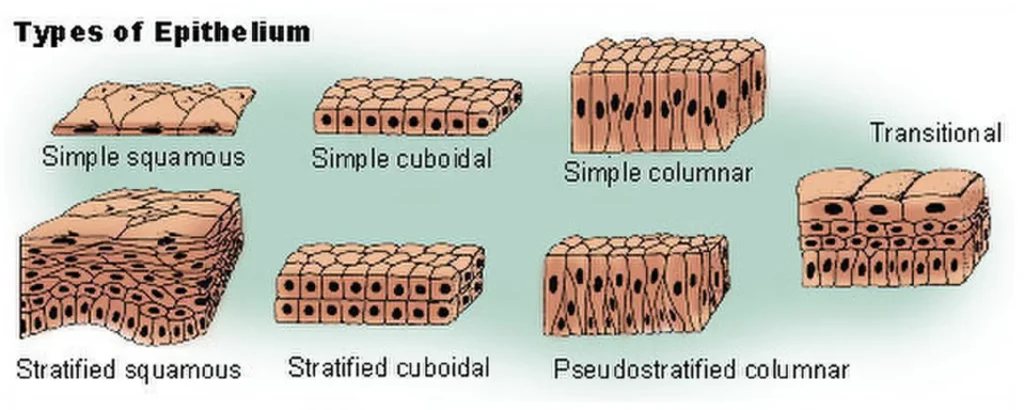
Single layer of cells resting on a basement membrane.
a. Simple Squamous Tissue
Large, flattened cells fitting like tiles on a floor (also known as Pavement Epithelium).
- Locations:
- Walls of blood vessels and lymph vessels (Endothelium).
- Lining of body cavity walls / Coelem (Mesothelium — cells are wavy, called Tesselated Epithelium).
- Lining of lung alveoli (Pneumocytes).
- Bowman’s capsule and Loop of Henle in Nephron.
- Functions: Exchange of materials between blood capillary and tissue fluid, gas exchange, and ultra-filtration.
b. Simple Cuboidal Epithelium
Single layer of cube-like cells with round central nucleus.
- Locations: PCT of nephron, ducts of glands, thyroid follicles, ovaries & testes (Germinal epithelium).
- Special Surface: Surface may have microvilli (Brush Border) to increase surface area for reabsorption.
- Functions: Reabsorption, secretion, and excretion.
c. Simple Columnar Epithelium
Tall, pillar-like cells with elongated nuclei located at the base.
- Locations: Lining of Stomach and Intestine.
- Special Cells: Contains Goblet cells (Granular cells) that secrete mucus in the alimentary canal.
- Functions: Secretion and absorption.
4. Ciliated Epithelium
Cuboidal or Columnar cells bearing hair-like cilia on their free surface.
- Cuboidal Ciliated: Found in Bronchioles (moves trapped mucus particles out of pharynx).
- Columnar Ciliated: Found in Fallopian tubes (facilitates movement of ova).
- Function: Transportation/movement of particles in a specific direction.
5. Pseudostratified Epithelium
Single layer of cells that appears multi-layered (stratified) because nuclei are located at different levels (contains long cells + short round-nucleated cells).
- Locations: Male urethra, Parotid gland duct, Trachea, and Bronchi.
6. Glandular Epithelium
Specialized cuboidal or columnar cells adapted primarily for secretion.
Classification by Cell Count:
- Unicellular Glands: Single isolated secretory cells. Example: Goblet cell (Mucocyte).
- Multicellular Glands: Clusters of cells forming duct and secretory units. Example: Salivary, Sweat, Gastric, Sebaceous glands.
Classification by Mode of Secretion:
- Merocrine (Eccrine) Glands: Secretion released via exocytosis without cell damage (e.g., Pancreas, Salivary glands).
- Apocrine Glands: Apical portion of the cell is discharged along with secretion (e.g., Mammary glands, Axillary sweat glands).
- Holocrine Glands: Entire cell ruptures and dies to release secretory product (e.g., Sebaceous oil glands).
7. Compound Epithelium
Made of more than one layer of cells. Main function is protection against mechanical and chemical stress (plays little role in secretion or absorption).
a. Stratified Epithelium Types:
- Keratinized Stratified Squamous: Covered with dead keratin protein layer. Impervious to water and resistant to friction. Example: Epidermis of skin.
- Non-Keratinized Stratified Squamous: Provides wet protection. Examples: Buccal cavity, Eye cornea, Pharynx, Oesophagus, Vagina.
- Stratified Cuboidal: Examples: Large ducts of salivary and pancreatic glands.
b. Transitional Epithelium
Highly stretchable, multi-layered epithelium. Lacks a distinct basement membrane (LBA absent).
- Locations: Urinary Bladder and Ureters.
7. Cellular Junctions
Structures that connect adjacent cells or connect cells to the extracellular matrix.
- Tight Junction (Zona Occludens): Prevents leakage of substances across tissues.
- Desmosome (Macula Adherens): Acts as anchoring junction using tonofibrils (keratin filaments).
- Gap Junction: Facilitates rapid cellular communication and transfer of ions/molecules between cells.
- Hemidesmosome: Anchors epithelial cells to the underlying Basement Membrane.
- Interdigitation: Interlocking finger-like processes for exchange (e.g., Placenta).
B. Connective Tissue
Most abundant and widely distributed tissue in the animal body. Connects different organs and forms packaging around organs.
- Matrix: Non-living ground substance made of polysaccharide-protein complexes secreted by cells.
- 3 Components: Cells + Matrix + Fibers.
Types of Cells in Connective Tissue:
- Fibroblasts: Most abundant cells; secretes fibers:
- Collagen Fibers: White, strong, unbranched, inelastic.
- Elastin Fibers: Yellow, flexible, branched, elastic.
- Macrophages (Histiocytes / Clasmatocytes): Derived from Monocytes; perform phagocytosis.
- Plasma Cells: Derived from B-lymphocytes; “Cart-wheel” shaped nucleus; secretes antibodies during infection.
- Mast Cells: Similar to Basophils; secretes three important substances:
- Histamine: Vasodilator (increases blood vessel diameter); Bronchioconstrictor (active in asthma).
- Serotonin: Vasoconstrictor (decreases blood vessel diameter).
- Heparin: Natural anticoagulant (maintains liquid nature of blood).
Types of Connective Tissue:
1) Loose Connective Tissue:
Matrix more
cells + Fibres – less
cells & Fibres – lossely
a. Areolar Tissue
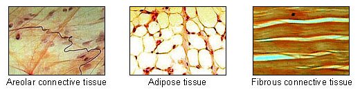
Most widely distributed connective tissue present beneath the skin. Serves as a support framework for epithelium.
- Joins skin to muscle and fills spaces inside organs around blood vessels and nerves.
- Contains Macrophages, Fibroblasts, and Mast cells.
- Function: Support, packaging, repair, and diffusion of materials.
b. Adipose Tissue
Fat-storing tissue composed of specialized cells called Adipocytes (contains fat globules).
- Locations: Subcutaneous skin layer, around heart, kidney, and eyeball.
- Functions: Acts as a shock absorber, fat reservoir, and insulator.
- Adipose tissue : Fat cells
Adipocytes:
Types of Adipocytes (White vs. Brown Fat)
a. White Adipose Tissue
- Monocular (single large central fat droplet).
- Contains fewer mitochondria.
- Functions as subcutaneous insulation, shock absorber, and reserve food storage.
- Cannot be easily removed from the body after exercise.
b. Brown Adipose Tissue
- Multilocular (several small fat droplets).
- Contains abundant mitochondria and cytochrome pigments giving it a brown color.
- Found in hibernating mammals and newborn babies.
- Generates heat to prevent shivering in newborns (never used as a food substrate).
2) Dense Connective Tissue
a. Dense Regular Connective Tissue
Collagen fibers are arranged in parallel rows between many parallel bundles.
- Tendon: Connects Bone to Muscle. Made of strong, inelastic white collagen fibers.
- Ligament: Connects Bone to Bone. Made of elastic yellow fibers with few collagen fibers.
*Sprain is caused due to excessive pulling/stretching of ligaments.
b. Dense Irregular Connective Tissue
Fibers and fibroblasts are oriented differently in various directions. Example: Skin dermis.
3) Specialized Connective Tissue: Cartilage
Harder than dense connective tissue but softer than bone. Matrix is solid, pliable, and resists compression.
- Cartilage Cells: Chondrocytes (enclosed in fluid-filled cavities called Lacunae, 2-4 cells per lacuna).
- Precursor Cells: Chondroblasts. Cartilage-destroying cells: Chondroclasts.
- Protein
Cartilage Types:
Matrix contains Chondrin protein. Cells are Chondrocytes inside fluid spaces called Lacunae.
- Hyaline: Articular joint surface, Nasal septum, Tracheal C-rings.
- White Fibrous: Strongest cartilage; Intervertebral disc, Pubic symphysis.
- Yellow Elastic: Tip of nose, Ear pinna, Epiglottis.
- Calcified: Head of long bones.
Bone Histology & Haversian System:
- Matrix: ~70% Inorganic + 30% Organic (Protein = Ossein).
- Bone Cells: Osteoblast (forming), Osteocyte (mature in lacuna), Osteoclast (destroying).
- Haversian System (Osteon): Longitudinal Haversian canals connected by transverse Volkmann’s canals. Characteristic feature of long mammalian bones.
C) Muscular Tissue:
Help in all movement and locomotion
All locomotion are movement but al movement are not locomotion
Types of Muscles:
- Skeletal Muscle: Cylindrical, striated, multinucleated, voluntary, fast contraction, fatigues easily.
- Smooth (Visceral) Muscle: Spindle-shaped, non-striated, uninucleated, involuntary, slow peristaltic action.
- Single-unit: Whole organ contracts together (Stomach, Uterus).
- Multi-unit: Contracts at specific areas (Blood vessels, hair root muscle).
- Cardiac Muscle: Cylindrical, striated, branched, involuntary, never fatigues; contains Intercalated Discs.
Sarcomere (Muscle Functional Unit):
- Segment between two successive Z-lines ($Z \to Z = 1 \text{ Sarcomere}$).
- A-Band: Dark band containing Myosin & Actin.
- I-Band: Light band containing only Actin.
- H-Zone: Center of A-band without Actin overlap; divided by M-line.
D) Nervous Tissue:
Conduction of impulse and control and coordination
Nervous Tissue & Neurons:
- Origin: Ectodermal (except Microglia = Mesodermal).
- Neuron: Contains Soma/Cell body with Nissl’s granules, Axon, and Synaptic Knobs.
- Myelin Sheath: Schwann cells (in PNS) and Oligodendrocytes (in CNS) increase impulse speed.
- Neuroglia Cells: Supporting non-neuronal cells ($>50\%$ volume of neural tissue).
Types of Neurons:
- Unipolar: 1 Axon only (Embryo).
- Bipolar: 1 Axon + 1 Dendrite (Retina of eye, Olfactory).
- Multipolar: 1 Axon + Many Dendrites (Cerebral cortex).
- Pseudounipolar: Bipolar looking unipolar (Dorsal root ganglion).