
Human Health and Diseases
1. AIDS (Acquired Immuno Deficiency Syndrome) & HIV Mechanics
A. Overview & Virus Structure
- Causative Agent: Human Immunodeficiency Virus (HIV) — a Retrovirus containing single-stranded RNA (ssRNA) enclosed within a protein envelope.• HIV-I: World-wide & highly common.• HIV-II: Rare & restricted mainly to African region.
- Structural Components:• Envelope glycoproteins: GP120 & GP41.• Phospholipid bilayer membrane $+$ Matrix protein ($P17$) $+$ Inner Capsid ($P24$).
• Genome: 2 identical copies of single-stranded RNA (ssRNA).
• Enzymes: Reverse Transcriptase (RNA $\to$ DNA), Integrase (integrates viral DNA into host genome), and Protease.
B. Replication Cycle & Mode of Action
- HIV enters Macrophages (via $CD_4$ receptors) where viral RNA replicates to form viral DNA via Reverse Transcriptase. Macrophages continue producing virus and act as an “HIV Factory”.
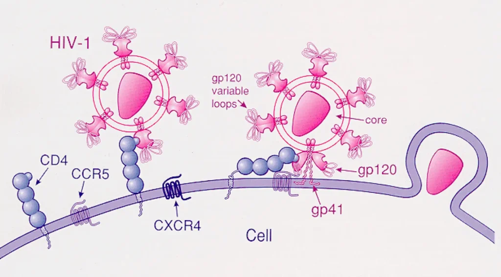
- HIV continuously enters Helper T-Lymphocytes ($T_H$ cells), replicates, and destroys them. When $T_H$ count drops below $200/\text{mm}^3$ of blood, full-blown AIDS manifests.
- Opportunistic Infections: Patient becomes vulnerable to pathogens like Toxoplasma, Mycobacterium tuberculosis, fungal infections, Kaposi’s Sarcoma, and Cytomegalovirus (CMV).
C. Transmission, Incubation, Diagnosis & Treatment
- Transmission: Unprotected sexual contact with infected individual, transfusion of contaminated blood/plasma, sharing infected needles/razors/tattooing, vertical transmission from mother to child via placenta. Does NOT spread by hugging, shaking hands, or social contact.
- Incubation Period: Time lag between infection and appearance of symptoms (usually 6 months to 10 years).
- Diagnostics: Screening test = ELISA (Enzyme-Linked Immunosorbent Assay based on Ag-Ab interaction). Confirmatory test = Western Blotting. Early detection via PCR.
- Treatment: No complete cure. Treated with Antiretroviral drugs (e.g., Azidothymidine / Zidovudine) which only prolong life.
2. Cancer Biology, Carcinogens & Treatments
A. Cancer Characteristics (Loss of Contact Inhibition)
- Abnormal, uncontrolled proliferation of cells. Normal cells possess Contact Inhibition (contact with neighboring cells inhibits uncontrolled growth); cancer cells lose this property.
- Cancerous cells form a mass of cells called a Tumor / Neoplasm, which starves normal cells by competing for vital nutrients.
- Tumor Types:• Benign Tumors: Remain confined to original location; cause little damage; non-cancerous.• Malignant Tumors: Mass of proliferating neoplastic cells. Display Metastasis (cells slough off, travel through blood, and initiate new tumors at distant sites – most feared property of cancer).
B. Causes of Cancer (Carcinogens)
| Category | Agents / Mechanisms | Target / Example |
|---|---|---|
| Physical Carcinogens | Ionizing radiations (X-rays, Gamma rays) & Non-ionizing radiation (UV rays). | DNA damage, skin cancer. |
| Chemical Carcinogens | Chemical agents like tobacco smoke, mustard gas, dyes. | Tobacco smoke major cause of Lung Cancer. |
| Biological Carcinogens | Oncogenic Viruses (Oncoviruses possessing viral oncogenes) or activation of cellular Proto-oncogenes / c-onc in normal cells. | Converts normal cells to neoplastic state under specific conditions. |
C. Detection, Diagnosis & Treatment
- Diagnostic Techniques: Biopsy & Histopathological studies, Radiography (X-rays), Computed Tomography (CT scan – 3D imaging using X-rays), MRI (Magnetic Resonance Imaging – uses strong magnetic fields and non-ionizing radiation; safest and most accurate), Monoclonal Antibodies (e.g., Herceptin), and Molecular gene testing.
- Treatment Modalities:1. Surgery: Surgical excision of tumor mass.2. Radiotherapy: Tumor cells irradiated lethally taking care of surrounding normal tissue.
3. Chemotherapy: Anti-cancer drugs like Vinblastine and Vincristine (extracted from Vinca rosea / Catharanthus roseus). Side effects = Hair loss, Anaemia.
4. Immunotherapy: Patients given biological response modifiers such as $\alpha$-Interferon which activates immune system to destroy tumor cells.
3. Innate vs. Acquired Immunity & Lymphoid Organs
A. Innate Immunity (4 Non-Specific Barriers)
Non-specific defence mechanism present right from birth:
- 1. Physical Barrier: Skin (Stratum corneum) prevents entry; Mucus coating of respiratory, gastrointestinal, and urogenital tracts traps microbes.
- 2. Physiological Barrier: Acid ($\text{HCl}$) in stomach, Saliva in mouth, Lysozyme in tears, ear wax, and sweat glands.
- 3. Cellular Barrier: Phagocytic cells like PMNL-neutrophils, Monocytes, Macrophages, and Natural Killer (NK) cells.
- 4. Cytokine Barrier: Virus-infected cells secrete proteins called Interferons ($\alpha, \beta, \gamma$) which protect non-infected cells from further viral infection.
B. Acquired Immunity (Humoral vs. Cell-Mediated)
Pathogen-specific immunity acquired during lifetime characterized by Memory, Diversity, and Self vs. Non-Self discrimination:
- Humoral Immune Response (HI / Antibody-Mediated): Mediated by B-Lymphocytes in body fluids (blood/lymph). Produce B-plasma cells (secretes antibodies; short life 4-5 days) and B-memory cells.
- Cell-Mediated Immunity (CMI): Mediated by T-Lymphocytes.• $T_K$ (Cytotoxic T-cells): Direct killing of infected/foreign cells.• $T_H$ (Helper T-cells): Stimulate B-cells to produce antibodies.
• $T_S$ (Suppressor T-cells) & $T_M$ (Memory T-cells).
• Graft Rejection: Responsible for rejection of transplanted organs (kidney, heart, liver). Suppressed during transplants using immunosuppressants like Cyclosporin A.
C. Primary vs. Secondary Lymphoid Organs
- Primary Lymphoid Organs: Sites where immature lymphocytes differentiate into antigen-sensitive cells $\to$ Bone Marrow (origin & maturation of B & T cells) and Thymus (maturation of T cells; atrophies with age).
- Secondary Lymphoid Organs: Sites where lymphocytes interact with antigens, undergo proliferation, and become effector cells $\to$ Spleen, Lymph Nodes, Tonsils, Peyer’s Patches of small intestine, Appendix.• Spleen: Large bean-shaped organ trapping blood-borne microorganisms; acts as a reservoir of erythrocytes (RBC graveyard).• MALT (Mucosa-Associated Lymphoid Tissue): Constitutes about $50\%$ of total lymphoid tissue in human body (located within respiratory, digestive, and urogenital tracts).
4. Antibody Structure, Immunoglobulin Classes & Autoimmunity
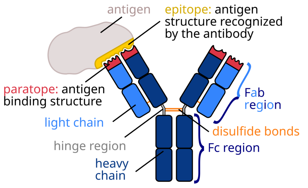
A. Structure of Antibody Molecule ($\text{H}_2\text{L}_2$)
- Y-shaped glycoprotein molecule consisting of 4 peptide chains: 2 Heavy (H) chains ($\approx 450\text{ aa}$) and 2 Light (L) chains ($\approx 220\text{ aa}$) held together by 16 Disulfide Bonds. Represented as $\mathbf{H}_2\mathbf{L}_2$.
- Paratope: Antigen-binding site located at the variable tip ($V_H / V_L$) of antibody; binds specifically to Epitope of antigen.
B. Classes of Immunoglobulins (Ig)
| Antibody Class | Molecular Structure | Key Features & Functions |
|---|---|---|
| IgA | Dimer (4 Paratopes) | 2nd most abundant; present in secretory fluids (Colostrum – 1st breast milk, saliva, tears). Provides natural passive immunity to infant. |
| IgG | Monomer (2 Paratopes) | Most abundant ($75\%$); ONLY antibody that crosses placenta from mother to fetus; primary driver of $2^\circ$ immune response. |
| IgM | Pentamer (10 Paratopes) | Largest antibody; first produced during primary ($1^\circ$) immune response. |
| IgE | Monomer (2 Paratopes) | Involved in Allergic Reactions & Hypersensitivity. Triggers Mast cells to release Histamine and Serotonin. |
| IgD | Monomer (2 Paratopes) | Membrane-bound on B-cell surface; acts as antigen receptor. |
C. Allergies & Autoimmune Diseases
- Allergies: Exaggerated hypersensitive immune response to environmental allergens (pollen, dust, animal dander, drugs). Mediated by IgE. Mast cells release vasodilators (Histamine & Serotonin). Symptoms treated with Anti-histamines, Adrenaline, and Steroids.
- Autoimmunity: Loss of self-tolerance where immune system attacks body’s own self-cells.• Examples: Myasthenia Gravis, Systemic Lupus Erythematosus (SLE), Grave’s Disease, Rheumatoid Arthritis, Vitiligo, Multiple Sclerosis, Type I Diabetes Mellitus.
5. Concepts of Health & Common Human Diseases (Part 1)
A. Early Concept of Health & William Harvey’s Disproof
- Historically, health was considered a state of body and mind with a balance of ‘humors’ (Good Humor Hypothesis by Hippocrates and Indian Ayurveda). Persons with ‘black bile’ were believed to belong to hot personality and have fevers.
- William Harvey (Discovery of Blood Circulation): Disproved the ‘Good Humor Hypothesis’ using experimental methods and thermometer measurements showing normal body temperatures in individuals with black bile.
- WHO Definition of Health: State of complete physical, mental, and social well-being, and not merely the absence of disease or infirmity. Affected by Genetic disorders, Lifestyle, and Infections.
B. Common Bacterial & Viral Diseases
- 1. Typhoid (Bacterial): Pathogen = Salmonella typhi.• Enters small intestine via contaminated food/water and migrates to organs via blood.• Symptoms: Sustained high fever ($39^\circ\text{C}$ to $40^\circ\text{C}$), weakness, stomach pain, constipation, headache, loss of appetite, intestinal perforation & death in severe cases.
• Diagnostic Test: Widal Test.
• Classic Case: Mary Mallon (“Typhoid Mary”), a cook who spread typhoid for several years through cooked food.
- 2. Pneumonia (Bacterial): Pathogens = Streptococcus pneumoniae & Haemophilus influenzae.• Infects fluid-filled alveoli of lungs, causing severe breathing difficulty.• Symptoms: Fever, chills, cough, headache; lips and finger nails may turn gray to bluish in color in severe cases. Spreads via droplets/aerosols.
- 3. Common Cold (Viral): Pathogen = Rhino Viruses.• Infects nose and respiratory passage, but DOES NOT INFECT LUNGS!• Symptoms: Nasal congestion, discharge, sore throat, hoarseness, cough, lasts $3\text{–}7\text{ days}$.
6. Protozoan Human Diseases (Malaria Species)
A. Malaria Overview & Plasmodium Species
- Caused by microscopic protozoan parasite Plasmodium (transmitted via female Anopheles mosquito vector).
- Major Human Pathogenic Species:• Plasmodium vivax: Causes Benign Tertian Malaria.• Plasmodium malariae: Causes Quartan Malaria.
• Plasmodium falciparum: Causes Malignant Malaria (most dangerous, severe, and can be fatal/brain damage!).