
Human Reproduction
1. Overview & Male Reproductive System
A. Sequence of Reproductive Events
- Gametogenesis: Formation of gametes (Sperm in males, Ova in females).
- Insemination: Transfer of sperms into female genital tract.
- Fertilization: Fusion of male and female gametes to form Zygote.
- Implantation: Attachment of Blastocyst to the uterine wall (Endometrium).
- Gestation: Embryonic/fetal development inside the womb (uterus).
- Parturition: Delivery of the fully developed fetus out of the mother’s body.
- Lactation: Production and release of milk by mammary glands to feed the newborn.
B. Male Reproductive System: Testes Anatomy
- Primary Sex Organ: Pair of Testes (Length: 4–5 cm, Width: 2–3 cm). Located extra-abdominally within the Scrotum to maintain temperature 2 to 2.5°C lower than normal body temperature for spermatogenesis.
- Testicular Descent: During the 7th month of fetal development, testes descend from abdomen to scrotum via the Inguinal canal under the influence of Testosterone.• Cryptorchidism: Failure of testes to descend into scrotum; leads to sterility.
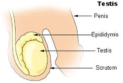
- Monorchidism: Presence of only one testis in scrotum (e.g., Ascaris).
- Temperature Regulation Muscles: Darter muscle (scrotal wall) & Cremaster muscle (spermatic cord) contract in winter (bring testes closer to body) and relax in summer (move testes away).
- Internal Structure: Each testis contains 250–300 testicular lobules. Each lobule has 1 to 3 highly coiled Seminiferous Tubules (structural unit of spermatogenesis).
- Duct Pathway:$$\text{Seminiferous Tubules} \to \text{Tubuli Recti} \to \text{Rete Testis} \to \text{Vasa Efferentia} \to \text{Epididymis} \to \text{Vas Deferens}$$
2. Accessory Glands, Sperm Structure & Female Organs
A. Male Accessory Glands
- Seminal Vesicles (Paired): Contributes 60–70% of semen volume. Secretes fructose (energy source for sperms), inositol, prostaglandins, citric acid, and fibrinogen (clotting protein).
- Prostate Gland (Single): Chestnut-shaped, lies below urinary bladder. Secretes alkaline fluid containing $\text{Ca}^{2+}$, citrate, and fibrinolysin to neutralize acidic vaginal environment. Has characteristic odor.
- Bulbourethral / Cowper’s Gland (Paired): Pea-sized, secretes alkaline viscous fluid for lubrication of penis during coitus.
• Per ejaculation volume: 2.5 to 3.5 mL (contains 200–300 million sperms).
• Normal Fertility Requirement: At least 60% sperms must have normal shape/size, and 40% must show vigorous motility.
• Oligospermia: Sperm count $< 20\text{ million/mL}$ (leads to infertility).
B. Structure of Mature Sperm
- Head: Contains haploid nucleus ($n=23$) capped by Acrosome (derived from Golgi body, contains hydrolytic enzymes / sperm lysins).
- Neck: Contains proximal centriole (initiates cleavage in zygote) and distal centriole (gives rise to axial filament).
- Middle Piece: Packed with spiral mitochondria (Nebenkern) that provide energy (ATP) for sperm motility.
- Tail: Long axial filament facilitating flagellar movement toward the ovum.
C. Female Reproductive System Overview
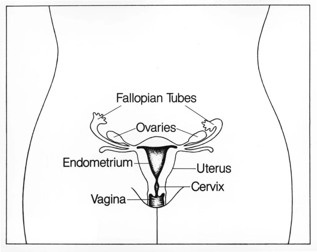
- Primary Sex Organ: Pair of Ovaries (produce ova and steroid hormones: Estrogen & Progesterone).
- Accessory Ducts: Fallopian tubes (Oviducts), Uterus (Womb), Cervix, Vagina.
- Fallopian Tube Divisions (10–12 cm long):$$\text{Infundibulum (Fimbriae)} \to \text{Ampulla (Site of Fertilization)} \to \text{Isthmus}$$
3. Ovary Anatomy, Folliculogenesis & Uterus Layers
A. Ovarian Anatomy & Attachments
- Almond-shaped organs (2–4 cm length) attached to pelvic wall and uterus by ligaments:• Mesovarium / Broad Ligament: Attaches ovary to abdominal wall.• Ovarian Ligament: Connects ovary to uterus.
• Suspensory Ligament: Connects ovary to pelvic wall.
- Ovarian Stroma: Divided into outer Cortex (contains follicles) and inner Medulla (contains blood vessels, elastic fibers, and connective tissue).
B. Follicular Development Stages
- Primary Follicle: Primary oocyte surrounded by a single layer of granulosa cells.
- Secondary Follicle: Surrounded by multiple granulosa layers and a theca layer.
- Tertiary Follicle: Characterized by a fluid-filled cavity called the Antrum (containing Liquor folliculi). Theca differentiates into Theca interna (hormonal) and Theca externa.
- Graafian Follicle (Mature): Contains secondary oocyte arrested at Metaphase II, surrounded by Zona Pellucida and Corona Radiata (Cumulus oophorus). Ruptures during ovulation under LH surge to release secondary oocyte.
- Corpus Luteum: Remnant of ruptured Graafian follicle; acts as temporary endocrine gland secreting large amounts of Progesterone. Degenerates into Corpus Albicans (white scar) if fertilization does not occur.
C. Structure of Uterus (Womb)
Inverted pear-shaped organ consisting of Fundus, Body (Corpus), and Cervix. Uterine wall consists of 3 layers:
- Perimetrium: External thin membranous layer.
- Myometrium: Middle thick layer of smooth muscle; exhibits strong contractions during labor (parturition).
- Endometrium: Inner glandular vascular layer.• Stratum Functionalis: Outer temporary layer sloughed off during menstruation.• Stratum Basalis: Permanent inner layer that regenerates functionalis.
4. Mammary Gland & Gametogenesis
A. Mammary Gland Pathway
Paired modified sweat glands characteristic of female mammals. Contain glandular tissue and variable fat.
Duct Pathway:
Mammary Lobules (15–20) $\to$ Alveoli $\to$ Mammary Tubules $\to$ Mammary Duct $\to$ Mammary Ampula $\to$ Lactiferous Duct $\to$ Nipple
B. Spermatogenesis vs Oogenesis Comparison
| Feature | Spermatogenesis | Oogenesis |
|---|---|---|
| Site & Onset | Seminiferous tubules; Starts at Puberty. | Ovary; Initiated during Fetal/Embryonic stage. |
| Division Pattern | Equal cytokinesis $\to$ 4 functional sperms ($n$). | Unequal cytokinesis $\to$ 1 Ovum ($n$) + Polar bodies. |
| Arrest Stages | Continuous process without arrest. | Arrested at Prophase I (Diplotene) & Metaphase II. |
| End Process | Spermiogenesis (Spermatid $\to$ Sperm) & Spermiation. | Completed only upon sperm entry at fertilization. |
5. Menstrual Cycle & Hormonal Control
A. Phases of Menstrual Cycle (28–29 Days)
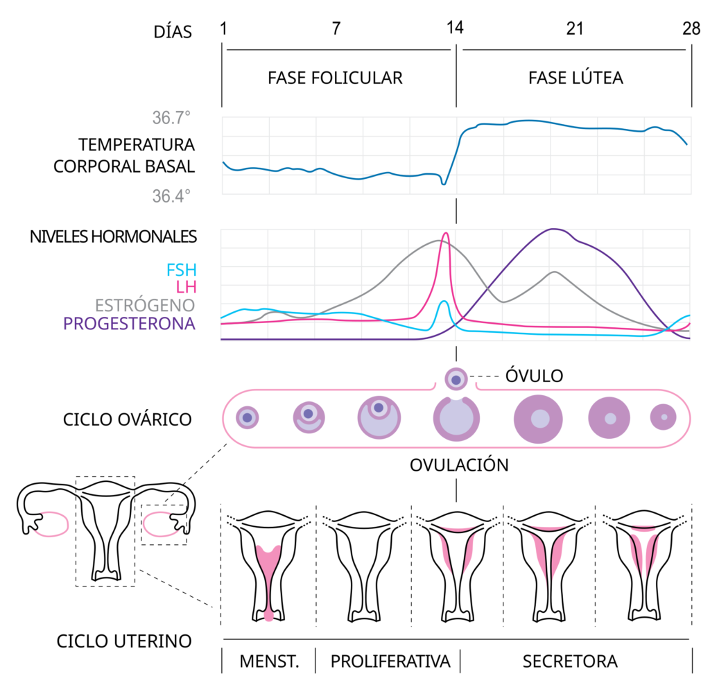
- 1. Menstrual Phase (Days 1–5): Breakdown of endometrial lining and blood vessels; flow lasts 3–5 days due to sudden drop in Progesterone levels.
- 2. Follicular / Proliferative Phase (Days 6–13): Primary follicles grow into Graafian follicles under FSH; Endometrium regenerates through proliferation stimulated by Estrogen.
- 3. Ovulatory Phase (Day 14): Peak levels of LH and FSH. Rapid secretion of LH leads to LH Surge, causing rupture of Graafian follicle and release of secondary oocyte (Ovulation).• Rule for Ovulation Day: $\text{Day of Ovulation} = \text{Total Cycle Length} – 14$. Luteal phase is fixed at 14 days.
- 4. Luteal / Secretory Phase (Days 15–28): Corpus luteum secretes high Progesterone to maintain endometrium for implantation. If fertilization fails, corpus luteum degenerates, causing menstruation.
B. Estrous Cycle (Non-Primate Mammals)
Occurs in cows, sheep, dogs, horses, rats. No menstrual bleeding; broken-down endometrium is reabsorbed. Females are receptive only during “Heat period” (High Estrogen).
6. Fertilization, Capacitation & Reactions
A. Sperm Capacitation & Chemoattraction
- Capacitation: Physiological conditioning of sperms in female genital tract (uterus/fallopian tube) lasting 6–7 hours. Involves removal of membrane cholesterol/glycoproteins and $\text{Ca}^{2+}$ influx, enabling hyperactive whiplash motility.
- Chemoattraction: Egg releases species-specific glycoprotein Fertilizin, which interacts with Antifertilizin (acidic protein) on sperm surface.
B. Acrosomal Reaction & Polyspermy Blocks
- Acrosomal Enzymes Released:• Hyaluronidase: Dissolves hyaluronic acid cementing corona radiata cells.• Corona Penetrating Enzyme (CPE): Dissolves corona radiata.
• Zona Lysin / Acrosin: Digests Zona Pellucida.
- Fast Block to Polyspermy: Binding of sperm to ZP3 receptor induces $\text{Na}^+$ influx, depolarizing the oocyte plasma membrane within seconds.
- Slow Block to Polyspermy (Cortical Reaction): $\text{Ca}^{2+}$ influx triggers exocytosis of cortical granules into perivitelline space, hardening Zona Pellucida and destroying ZP3 receptors.
7. Cleavage & Implantation
A. Cleavage Dynamics
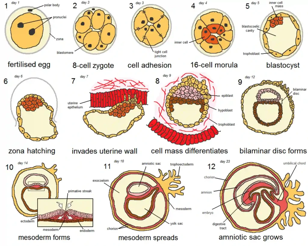
- Rapid mitotic divisions of zygote in Fallopian tube. First cleavage takes ~30 hours.
- Pattern: Holoblastic (complete division), unequal, and Rotational cleavage.
- Morula: Solid ball of 8–16 blastomeres with tight junctions. Overall size does not increase due to presence of intact Zona Pellucida.
- Blastocyst / Blastula: Formed on Day 4–5. Consists of outer Trophoblast layer (forms extra-embryonic membranes/placenta) and Inner Cell Mass (ICM / Embryoblast) (forms complete embryo body). Contains fluid cavity called Blastocoel.
B. Zona Hatching & Implantation
- Zona Hatching: Trophoblast secretes lytic enzymes to digest Zona Pellucida, allowing sticky blastocyst to hatch out.
- Implantation: Attachment of blastocyst to uterine endometrium on 6th–7th day after fertilization.
- Ectopic Pregnancy: Implantation occurring at any site other than uterus (e.g., Tubal pregnancy in fallopian tube).
- Decidua Layers:• Decidua Basalis: Endometrium between blastocyst and myometrium (forms maternal part of placenta).• Decidua Capsularis: Endometrial layer covering blastocyst.
• Decidua Parietalis: Remaining lining of uterine cavity.
8. Gastrulation & Extra-Embryonic Membranes
A. Gastrulation (Germ Layer Formation)
Process of morphogenetic cell movements transforming monoblastic blastula into triploblastic gastrula containing three primary germ layers.
- Inner cell mass differentiates into Epiblast (forms 3 germ layers) and Hypoblast. Formation of Primitive Streak marks commencement of gastrulation.
B. Fate of Primary Germ Layers
| Germ Layer | Derivatives / Derived Organs |
|---|---|
| Ectoderm | Epidermis of skin, Central & Peripheral Nervous System, Eye (lens/retina), Internal Ear, Pituitary Gland, Pineal Gland, Adrenal Medulla. |
| Mesoderm | Dermis of skin, Muscles, Connective tissues, Endoskeleton (Bones/Cartilage), Blood & Blood vessels, Heart, Kidneys, Gonads, Adrenal Cortex, Spleen. |
| Endoderm | Gut lining (GI tract), Visceral organs (Lungs, Trachea, Liver, Pancreas, Thyroid, Parathyroid, Thymus), Urinary bladder lining, Eustachian tube. |
C. Extra-Embryonic Membranes
- Amnion (Inner): Fills with Amniotic Fluid; prevents desiccation, acts as shock absorber.
- Chorion (Outer): Forms fetal portion of placenta; secretes hCG.
- Yolk Sac: Site of early blood cell formation (Haemopoiesis).
- Allantois: Forms blood vessels of umbilical cord; excretory/respiratory in lower vertebrates.
9. Placenta & Gestation Period
A. Placental Nature & Endocrine Hormones
Structural and functional unit between developing embryo/fetus and maternal body. Human placenta is Deciduate, Hemochorial, Discoidal, and Choriosantoic.
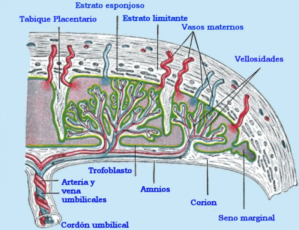
- Hormones Secreted by Placenta:• hCG (Human Chorionic Gonadotropin): Maintains Corpus Luteum to continue progesterone release. Presence in urine confirms pregnancy (Gravindex / Pregnancy Test).• hPL / hCS (Human Placental Lactogen / Somatomammotropin): Prepares mammary glands for lactation and regulates maternal glucose metabolism.
• Estrogen & Progesterone: Maintain uterine lining and prevent menstruation.
• hCT & hCC: Human Chorionic Thyrotropin and Corticotropin.
• Relaxin: Secreted by Corpus Luteum of ovary toward end of gestation; loosens pubic symphysis ligament for easier delivery.
B. Human Fetal Milestone Timeline
- Gestation Period: 266 days from fertilization (280 days / 40 weeks from Last Menstrual Period – LMP).
- 1st Month: Heart is formed (first sign of growing fetus is heart sound via stethoscope).
- 2nd Month: Limbs and digits develop.
- 3rd Month (End of 1st Trimester): Major organ systems formed, including external genital organs.
- 5th Month: First fetal movements felt by mother and appearance of fine hair on head.
- 6th Month (End of 2nd Trimester): Body covered with fine hair, eye-lids separate, and eye-lashes formed.
10. Parturition & Lactation
A. Neuroendocrine Reflex of Parturition
Process of child birth induced by a complex neuroendocrine mechanism.
- Fully developed fetus and placenta originate signals causing mild uterine contractions called Foetal Ejection Reflex.
- Triggers release of Oxytocin from maternal posterior pituitary gland.
- Oxytocin acts on myometrium, stimulating stronger uterine contractions, which further stimulates more oxytocin secretion (Positive Feedback Loop).
- Leads to cervical dilation and expulsion of baby through birth canal.
- Within 15–20 minutes after delivery, placenta and umbilical cord remnants are expelled (Afterbirth).
Post-delivery, fetal respiration switches from placental to pulmonary. Gaseous hormone Nitric Oxide (NO) acts as a vasodilator to dilate pulmonary blood vessels in newborn. Cortisol regulates timing of birth.
B. Lactation & Colostrum Immunity
- Hormone Control:• Prolactin (Anterior Pituitary): Stimulates milk synthesis and secretion.• Oxytocin (Posterior Pituitary): Stimulates milk ejection / let-down reflex.
- Colostrum: Yellowish thick fluid produced during initial few days of lactation. Rich in proteins, minerals, and rich in IgA antibodies providing essential Passive Immunity to newborn baby.