
Neural Control and Coordination
1. Neuron Structure, Myelination & Impulse Conduction
A. Neuron (Structural & Functional Unit)
- Excitable and conductive cells. Neural system originated first in Coelenterata (Cnidaria – e.g., Hydra) as a basic nerve net.
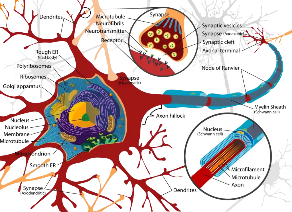
- Cyton / Cell Body: Contains cytoplasm, nucleus, and Nissl’s Granules (clumps of Rough Endoplasmic Reticulum and ribosomes for protein synthesis; absent in axon).
- Dendrites: Short, branched processes that receive impulses and transmit toward cell body (centripetal/unidirectional).
- Axon: Long single process transmitting impulses away from cell body (centrifugal). Originates from Axon Hillock (most sensitive part/trigger zone).• Ends in knob-like structures called Synaptic Knobs containing synaptic vesicles loaded with neurotransmitters.
B. Myelination & Saltatory Conduction
- Myelin Sheath: Insulating lipid layer around axon.• In PNS: Formed by Schwann Cells (myelinated cranial & spinal nerves).
• In CNS: Formed by Oligodendrocytes (white matter of brain & spinal cord).
- Nodes of Ranvier: Uninsulated gaps along myelinated axon lacking myelin sheath.
- Saltatory Conduction: Impulse jumps from one Node of Ranvier to another. Increases conduction speed drastically, requiring less time, energy, and ATP.
- Factors Increasing Conduction Speed: Myelination $>$ Axon Diameter (Thicker axon = faster speed; e.g., Giant Squid axon) $>$ Higher Temperature.
C. Types of Neurons Based on Structure
| Neuron Type | Structural Description | Location / Examples |
|---|---|---|
| Apolar / Non-polar | No distinct axon or dendrite; primitive network. | Hydra (Cnidarians). |
| Unipolar | Cell body with only one single process (axon). | Embryonic developmental stages. |
| Bipolar | One axon and one dendrite extending from cell body. | Retina of eye, Olfactory/Nasal epithelium. |
| Multipolar | One axon and two or more dendrites. | Cerebral Cortex, Most neurons in CNS/PNS. |
| Pseudounipolar | Single process arises from cyton and splits into two. | Dorsal root ganglion of spinal cord. |
2. Electrophysiology of Nerve Impulse & Synapse
A. Phases of Action Potential
| Phase | Membrane Potential | Ion Channel & Permeability State |
|---|---|---|
| 1. Resting State (Polarized) | $-70\text{ mV}$ | Axolemma more permeable to $K^+$ than $Na^+$. Active $\text{Na}^+/\text{K}^+$ Pump pumps $3\text{ Na}^+$ OUT for every $2\text{ K}^+$ IN using ATP. Outside positive, inside negative (negatively charged proteins in axoplasm). |
| 2. Depolarization (Action Potential) | Threshold: $-55\text{ to } -65\text{ mV}$ $\to$ Peak: $+35\text{ mV}$ | Threshold stimulus opens Voltage-Gated $\text{Na}^+$ Channels. Rapid influx of $\text{Na}^+$ ions turns inside positive and outside negative. Generates Action Potential (Nerve Impulse). |
| 3. Repolarization | Returns from $+35\text{ mV} \to -70\text{ mV}$ | $\text{Na}^+$ channels close; Voltage-Gated $\text{K}^+$ Channels open. Rapid efflux of $K^+$ ions restores negative charge inside. |
| 4. Hyperpolarization | Drops below resting level ($\approx -90\text{ mV}$) | Slow closure of $K^+$ channels allows excess $K^+$ efflux. Restored back to $-70\text{ mV}$ by $\text{Na}^+/\text{K}^+$ pump during refractory period. |
B. Synaptic Transmission Mechanics
- 1. Electrical Synapse: Membranes of pre- and post-synaptic neurons in close proximity (gap junction $\le 2\text{ nm}$). Direct ionic current flow; faster transmission; rare in humans.
- 2. Chemical Synapse: Separated by fluid-filled Synaptic Cleft ($> 20\text{ nm}$). Requires neurotransmitters. Has a minor delay ($\approx 1\text{ ms}$).
- Mechanism at Chemical Synapse:$$\text{Impulse arrives at synaptic knob} \to \text{Opens Voltage-Gated Ca}^{2+} \text{ channels} \to \text{Ca}^{2+} \text{ influx} \to \text{Exocytosis of neurotransmitter vesicles} \to \text{Release into cleft} \to \text{Binds post-synaptic receptors} \to \text{Opens ion channels}$$
3. Neurotransmitters, Nervous System & Meninges
A. Neurotransmitter Types
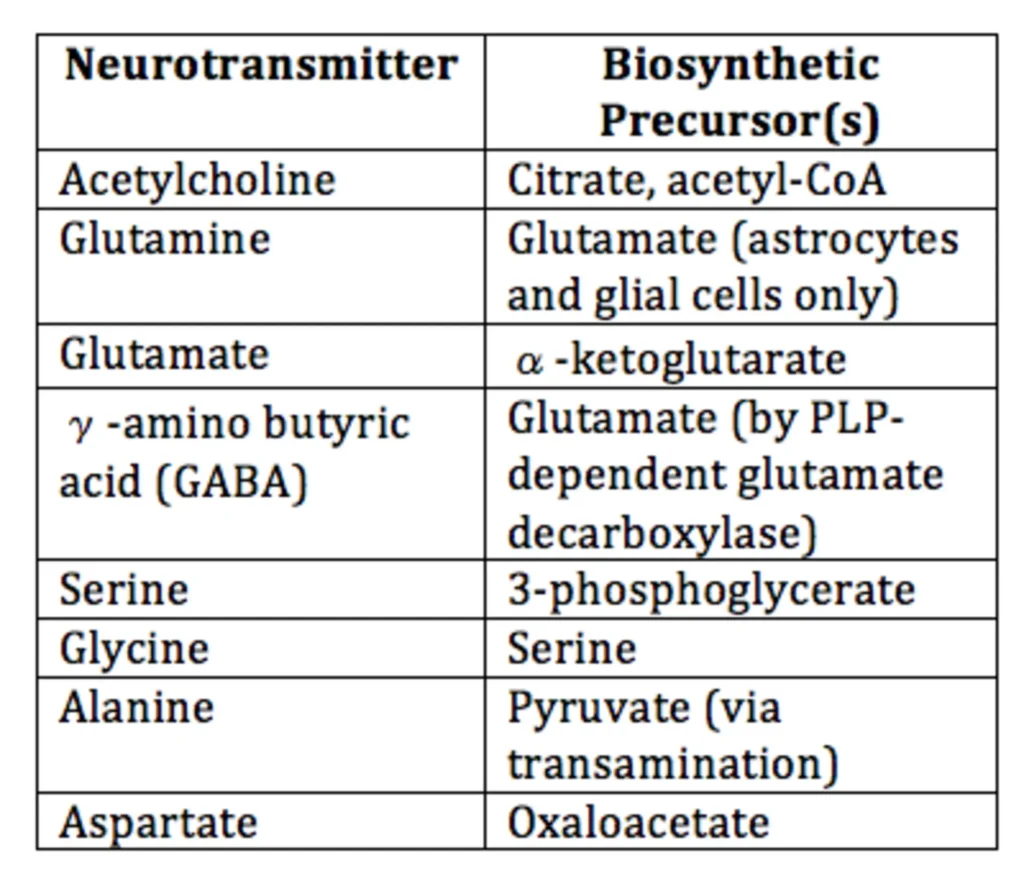
- 1. Excitatory / Stimulatory: Causes depolarization by opening $\text{Na}^+$ channels.• Acetylcholine (ACh): Broken down by Acetylcholinesterase into acetic acid and choline. Important for memory; low ACh causes Alzheimer’s Disease.
• Noradrenaline / Adrenaline & Glutamate.
- 2. Inhibitory: Causes hyperpolarization by opening $K^+$ or $Cl^-$ channels.• Dopamine: Deficiency in basal ganglia causes Parkinson’s Disease (tremors/shaky movements); excess causes Schizophrenia (hallucinations).
• GABA (Gamma-Amino Butyric Acid) & Glycine.
B. Divisions of Human Nervous System
- Central Nervous System (CNS): Brain $+$ Spinal Cord (Control and processing centre).
- Peripheral Nervous System (PNS): All nerves connecting CNS to body parts.• Cranial Nerves: 12 pairs in humans.
• Spinal Nerves: 31 pairs in humans.
• Somatic Nervous System (SNS): Relays impulses from CNS to voluntary skeletal muscles.
• Autonomic Nervous System (ANS): Relays impulses from CNS to involuntary smooth/cardiac muscles.
↳ Sympathetic: Emergency system ($3F$ – Fight, Flight, Fright).
↳ Parasympathetic: Rest and Digest system.
C. Cranial Meninges & CSF
Protected by skull bone and 3 connective tissue membranes (Meninges):
- Duramater (Outer): Tough, fibrous, hard, non-vascular layer. Subdural space lies beneath it.
- Arachnoid (Middle): Thin, delicate web-like layer (present only in mammals). Subarachnoid space lies beneath it.
- Piamater (Inner): Highly vascularized membrane in direct contact with brain tissue.
- Cerebrospinal Fluid (CSF): Secreted by choroid plexus in brain ventricles; fills subarachnoid space and central canal. Acts as a shock absorber and cushion protecting brain against mechanical injury.
4. Brain Anatomy, Limbic System & Functional Centers
A. Forebrain (Prosencephalon)
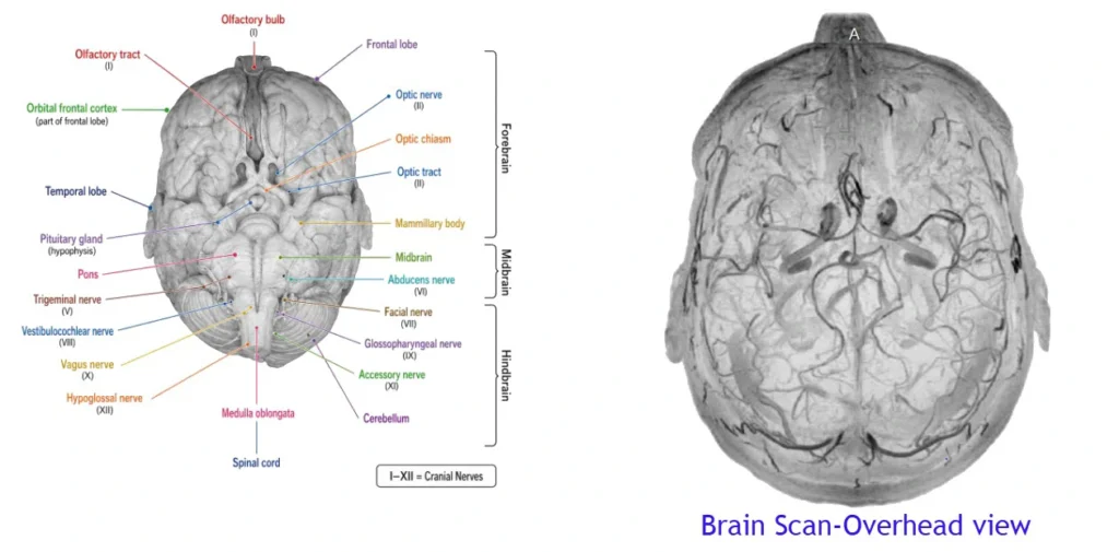
- 1. Cerebrum: Largest part of brain. Divided into Left and Right Cerebral Hemispheres joined by a thick tract of transverse nerve fibers called Corpus Callosum (characteristic of mammals).• Outer Cortex (Grey Matter): Contains neuron cytons and unmyelinated axons; folded into Gyri (ridges) and Sulci (grooves) to increase surface area.
• Inner Matter (White Matter): Contains myelinated axon tracts.
• Cerebral Cortical Areas: Sensory areas, Motor areas, and Association Areas (neither purely sensory nor motor; responsible for complex functions like memory, communication, and intersensory associations).
- 2. Diencephalon:• Thalamus: Major relay center for sensory and motor signaling.
• Hypothalamus: Lies at base of thalamus. Contains centers controlling thermoregulation ($37^\circ\text{C}$ body temp), hunger, thirst, satiety, and hormone secretion.
- 3. Olfactory Lobes: Center for sense of smell.
B. Limbic System (“Emotional Brain”)
Inner parts of cerebral hemispheres and associated deep structures (Amygdala, Hippocampus, Hypothalamus):
- Amygdala: Almond-shaped structure; converts fear into aggression/anger; essential for survival reactions.
- Hippocampus: Converts short-term memory into long-term memory; aids memory recall.
- Nucleus Accumbens: Activates reward and pleasure pathway.
C. Midbrain & Hindbrain Anatomy
- Midbrain (Mesencephalon): Located between thalamus/hypothalamus and pons.• Dorsal surface features 4 rounded swellings/lobes called Corpora Quadrigemina (Superior colliculi for visual reflexes; Inferior colliculi for auditory reflexes).
• Traversed by Cerebral Aqueduct (Aqueduct of Sylvius) connecting $3^{\text{rd}}$ and $4^{\text{th}}$ ventricles.
• Contains Cerebral Peduncles connecting hindbrain to forebrain.
- Hindbrain (Rhombencephalon):• Cerebellum: Second largest part of brain. Highly folded surface with “Tree of Life” pattern (Arbor Vitae in white matter) and Purkinje cells. Controls posture, equilibrium, motor coordination, and learned motor skills (typing, running).
• Pons Varolii: Fiber tracts interconnecting different brain regions. Contains Pneumotaxic Centre regulating breathing rate.
• Medulla Oblongata: Connected to spinal cord. Contains involuntary centers regulating Cardiovascular reflexes, Respiration (Respiratory Rhythm Centre), Swallowing, Gastric secretions, and Vomiting.
- Brain Stem: Formed by Midbrain $+$ Pons Varolii $+$ Medulla Oblongata (connects brain to spinal cord).