Locomotion and Movement
1. Types of Movements & Muscle Classification
A. Movement vs. Locomotion
- Movement: Change in position of any body part at the same place.
- Locomotion: Voluntary movement resulting in change of place/location.
- Key Principle: All locomotions are movements, but all movements are not locomotion. Plants show movement but no locomotion. Animals show both (except sponges and few coelenterates).
B. Types of Movements in Human Body
- 1. Amoeboid Movement: Exhibited by Macrophages and Neutrophils (WBCs) via pseudopodia formed by cytoplasmic streaming (actin microfilaments).
- 2. Ciliary & Flagellar Movement:• Ciliary: Trachea (removes dust/foreign particles), Fallopian tube (propels ova), Paramecium.
• Flagellar: Human Sperms, Choanocytes in Sponges canal system.
- 3. Muscular Movement: Movement of limbs, jaws, tongue, etc., supported by skeletal system.
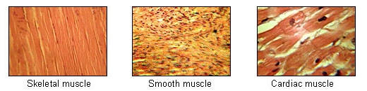
C. Types of Muscle Tissues
| Feature | Skeletal Muscle | Smooth / Visceral Muscle | Cardiac Muscle |
|---|---|---|---|
| Shape & Structure | Cylindrical, Unbranched. | Fusiform (spindle-shaped), Unbranched. | Cylindrical, Branched. |
| Nucleus & Striation | Multinucleated (Syncytium); Striated (Dark & Light bands). | Uninucleated; Non-striated (Smooth). | Uninucleated; Faintly striated with Intercalated discs. |
| Control & Location | Voluntary; Attached to skeletal bones. | Involuntary; Internal visceral organs. | Involuntary; Wall of Heart. |
D. Ultrastructure of Muscle & Sarcomere
- Organization: Muscle $\to$ Muscle Bundles (Fascicles held by collagenous Fascia) $\to$ Muscle Fibres (cells) $\to$ Myofibrils $\to$ Myofilaments (Thick Myosin & Thin Actin).
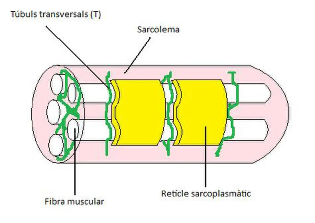
- Muscle Fibre Cell: Lined by Sarcolemma (plasma membrane) enclosing Sarcoplasm and storehouse of $Ca^{2+}$ called Sarcoplasmic Reticulum (SR).
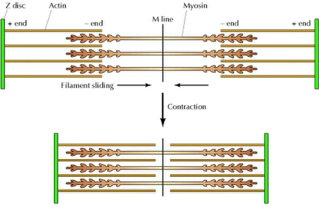
- Sarcomere (Functional Unit of Contraction): Region between two consecutive Z-lines.
- • A-Band (Anisotropic): Dark band containing thick Myosin filaments (and overlapping Actin). Length = $1.5\ \mu\text{m}$.
• I-Band (Isotropic): Light band containing only thin Actin filaments, bisected by Z-line.
• H-Zone (Hensen’s Zone): Central part of A-band not overlapped by thin filaments. Bisected by M-line.
• Formula: $\mathbf{1\text{ Sarcomere} = 1\text{ A-Band} + 2 \times (\frac{1}{2}\text{ I-Band})}$.
2. Contractile Proteins & Sliding Filament Theory
A. Structure of Contractile Proteins
- 1. Thick Filament (Myosin): Polymer of monomeric Meromyosins.• Heavy Meromyosin (HMM): Globular Head $+$ Short Arm (Cross arm); contains active $ATPase$ enzyme, ATP binding site, and Actin binding site.
• Light Meromyosin (LMM): Fibrous Tail region.
- 2. Thin Filament (Actin, Tropomyosin, Troponin):
- • F-Actin (Fibrous): Polymer of double-stranded helical G-Actin (Globular) monomers ($Mg^{2+}$ dependent).
• Tropomyosin: Two fibrous strands running close to F-actin grooves.
• Troponin: Complex trimeric protein (TpT – binds tropomyosin, TpI – inhibits actin-myosin interaction, TpC – binds $Ca^{2+}$) resting on tropomyosin to mask myosin binding sites during relaxation.
B. Neuromuscular Junction & Contraction Cascade
- Nerve impulse reaches motor end-plate $\to$ Releases Neurotransmitter Acetylcholine (ACh) into synaptic cleft.
- ACh binds to receptors on sarcolemma $\to$ Generates action potential propagating through T-tubules to Sarcoplasmic Reticulum (SR).
- SR releases $\mathbf{Ca^{2+}}$ into sarcoplasm $\to$ $Ca^{2+}$ binds to Troponin-C (TpC).
- Conformational shift unmasks myosin-binding sites on actin filaments.
- Myosin head hydrolyzes ATP ($\text{ATP} \xrightarrow{ATPase} \text{ADP} + P_i$) $\to$ Binds to exposed actin site forming a Cross-Bridge.
- Power Stroke: Myosin pulls attached actin filament inward toward center of A-band (M-line).
- New ATP binds to myosin head $\to$ Cross-bridge breaks. Process repeats as long as $Ca^{2+}$ levels remain elevated.
C. Changes During Muscle Contraction
| Sarcomere Zone / Line | Status During Contraction |
|---|---|
| Sarcomere Length | Decreases (Shortens). |
| I-Band (Light Band) | Decreases (Shortens). |
| H-Zone | Decreases / Disappears completely. |
| A-Band (Dark Band) | NO CHANGE (Retains original length). |
| Z-Lines | Pulled inwards toward M-line. |
3. Red vs. White Muscle Fibres & Skull Bones
A. Red vs. White Muscle Fibres
| Feature | Red Muscle Fibres (Type I) | White Muscle Fibres (Type II) |
|---|---|---|
| Myoglobin & Colour | High myoglobin; Reddish appearance. | Low myoglobin; Pale or whitish appearance. |
| Mitochondria & Respiration | Abundant mitochondria; Aerobic breakdown. | Few mitochondria; Anaerobic breakdown (Lactic acid accumulation). |
| Contraction Speed & Fatigue | Slow, sustained contraction; Does not fatigue easily. | Fast, rapid contraction; Fatigues quickly. |
| Sarcoplasmic Reticulum | Less sarcoplasmic reticulum. | High sarcoplasmic reticulum content. |
| Examples | Extensor muscles of back, Flight muscles of migratory birds. | Eyeball muscles, muscles of eyelids, short flight birds. |
B. Human Skeletal System Overview (206 Bones)
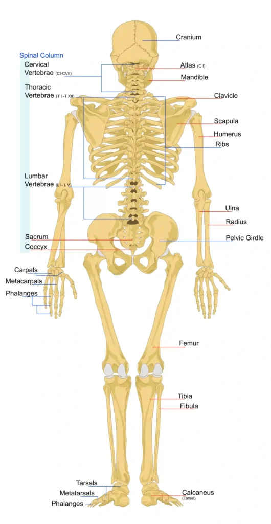
Divided into Axial Skeleton (80 bones) and Appendicular Skeleton (126 bones).
- Axial Skeleton (80): Skull (22) $+$ Hyoid (1) $+$ Ear Ossicles (6) $+$ Vertebral Column (26) $+$ Sternum (1) $+$ Ribs (24).
- 1. Skull Bones (22 Bones):• Cranium (8 Bones – Brain box): Paired: Parietal (2), Temporal (2). Unpaired: Frontal (1), Occipital (1), Sphenoid (1 – bat-shaped bone with Sella Turcica housing pituitary gland), Ethmoid (1).
• Human skull is Dicondylic (possesses 2 occipital condyles articulating with Atlas vertebra). Large opening at skull base = Foramen Magnum.
• Facial Bones (14 Bones): Nasal (2), Inferior Nasal Conchae (2), Lacrimal (2), Palatine (2), Vomer (1), Zygomatic (2 – cheek bones), Maxilla (2 – upper jaw), Mandible (1 – lower jaw; only movable bone of skull).
4. Vertebral Column, Ribs & Sternum
A. Ear Ossicles, Hyoid & Vertebral Column
- Ear Ossicles (6): Malleus (hammer-shaped), Incus (anvil-shaped), Stapes (stirrup-shaped – smallest bone of body). Mnemonic: MIS.
- Hyoid Bone (1): Single U-shaped bone at base of buccal cavity; does not articulate with any other bone.
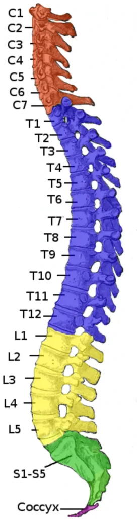
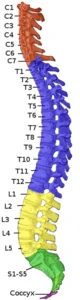
- Vertebral Column (26 Adult Vertebrae):• Formula: $\mathbf{C_7 T_{12} L_5 S_{(1)} C_{(1)}}$ (Sacral 5 fused into 1; Coccygeal 4 fused into 1).
• Cervical ($C_1\text{–}C_7$): $C_1 = \text{Atlas}$ (nodding ‘Yes’ joint with occipital condyles), $C_2 = \text{Axis}$ (pivot ‘No’ joint via odontoid process). All mammals have 7 cervical vertebrae (Exceptions: Sea cow & Sloths).
• Spinal Curvatures: Primary curves (Thoracic, Sacral – present at birth); Secondary curves (Cervical – forms at 3 months when holding head, Lumbar – forms at 6 months when sitting/walking).
B. Sternum & Rib Cage (24 Ribs)
Ribs are Bicephalic (possess two articulation surfaces on dorsal head end). Attached dorsally to thoracic vertebrae and ventrally to sternum via Hyaline Costal Cartilage.
- True Ribs / Vertebrosternal (Pairs 1 to 7): Directly attached to sternum via costal cartilage.
- False Ribs / Vertebrochondral (Pairs 8, 9, 10): Do not directly articulate with sternum; join the costal cartilage of the 7th rib.
- Floating Ribs / Vertebral (Pairs 11 & 12): Ventral ends remain unattached/free; protect kidneys.
- Rib Cage Assembly: Formed by Thoracic Vertebrae (dorsal), Ribs (lateral), and Sternum (ventral).
5. Appendicular Skeleton, Joints & Disorders
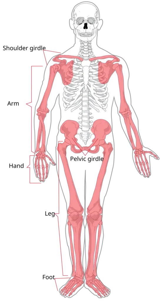
A. Appendicular Skeleton (126 Bones)
- Pectoral Girdle (4 Bones):• Scapula (2): Large triangular flat bone on dorsal thorax ($2^{\text{nd}}$ to $7^{\text{th}}$ rib). Features spine, acromion process, and Glenoid Cavity (articulates with head of humerus).
-
• Clavicle (2): Collar bone; long S-shaped bone with two articulations.
- Pelvic Girdle (2 Coxal / Hip Bones): Formed by fusion of Ilium, Ischium, and Pubis. Features deep cup cavity called Acetabulum (articulates with head of femur). Two halves meet ventrally at Pubic Symphysis (fibrocartilag