Human Excretion
1. Nitrogenous Wastes & Human Excretory Anatomy
A. Modes of Excretion & Nitrogenous Wastes
| Feature | Ammonotelism | Ureotelism | Uricotelism |
|---|---|---|---|
| Toxicity Level | Most Toxic | Moderately Toxic ($100,000\times$ less toxic than $\text{NH}_3$) | Least Toxic |
| Water Required | Maximum water required for excretion. | Moderate water required. | Minimum water (Excreted as semi-solid paste/pellet). |
| Energy / Excretion Site | No metabolic energy needed; simple diffusion via body surface/gills. No role of kidney. | Synthesized in Liver via Ornithine Cycle; filtered by Kidneys. | Most expensive metabolically (requires maximum ATP energy). |
| Examples | Bony fishes, aquatic amphibians (Tadpole), aquatic insects. | Cartilaginous fishes, adult amphibians, mammals. | Reptiles, Birds, Land Snails, Insects. |
B. Human Excretory System Components
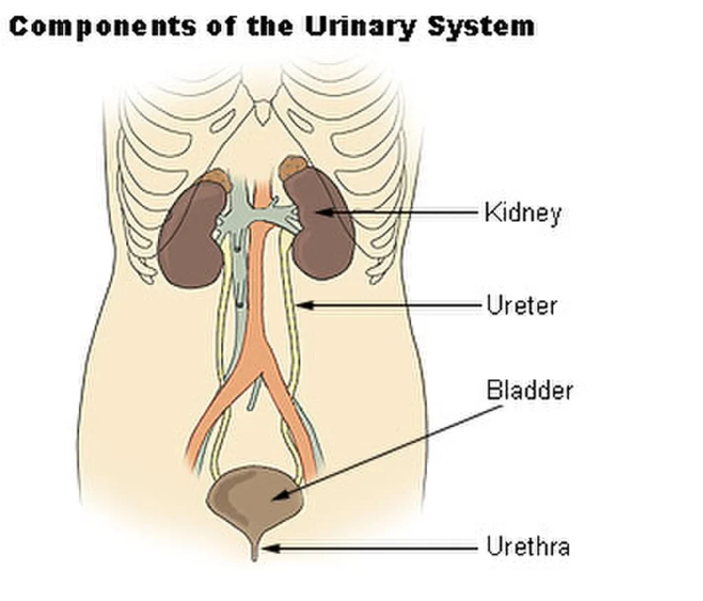
- 1. Kidneys (Pair): Reddish-brown, bean-shaped organs (Length: 10–12 cm, Width: 5–7 cm, Thickness: 2–3 cm, Weight: 120–170 g).• Positioned between $T_{12}$ and $L_3$ vertebrae attached to dorsal abdominal wall. Covered by peritoneum on ventral side only $\to$ Retroperitoneal / Metanephric Kidney.
• Outer Renal Cortex & Inner Renal Medulla (divided into medullary pyramids projecting into calyces). Cortex extending between pyramids forms Columns of Bertini.
- 2. Ureters (Pair): Hollow muscular tubes lined by stretchable Transitional Epithelium. Propels urine via peristaltic movements.
- 3. Urinary Bladder: Muscular sac containing smooth Detrusor Muscle (longitudinal-circular-longitudinal) and a triangular base called Trigone of Bladder. Stores up to 700–800 mL of urine.
- 4. Urethra: Canal guarded by Internal Urethral Sphincter (involuntary) and External Urethral Sphincter (voluntary).• Longer in males, shorter in females $\to$ Urinary Tract Infections (UTI) are more common in females.
2. Urea Cycle, Nephron Structure & Types
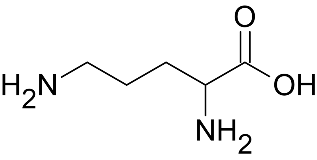
A. Urea / Krebs-Henseleit / Ornithine Cycle
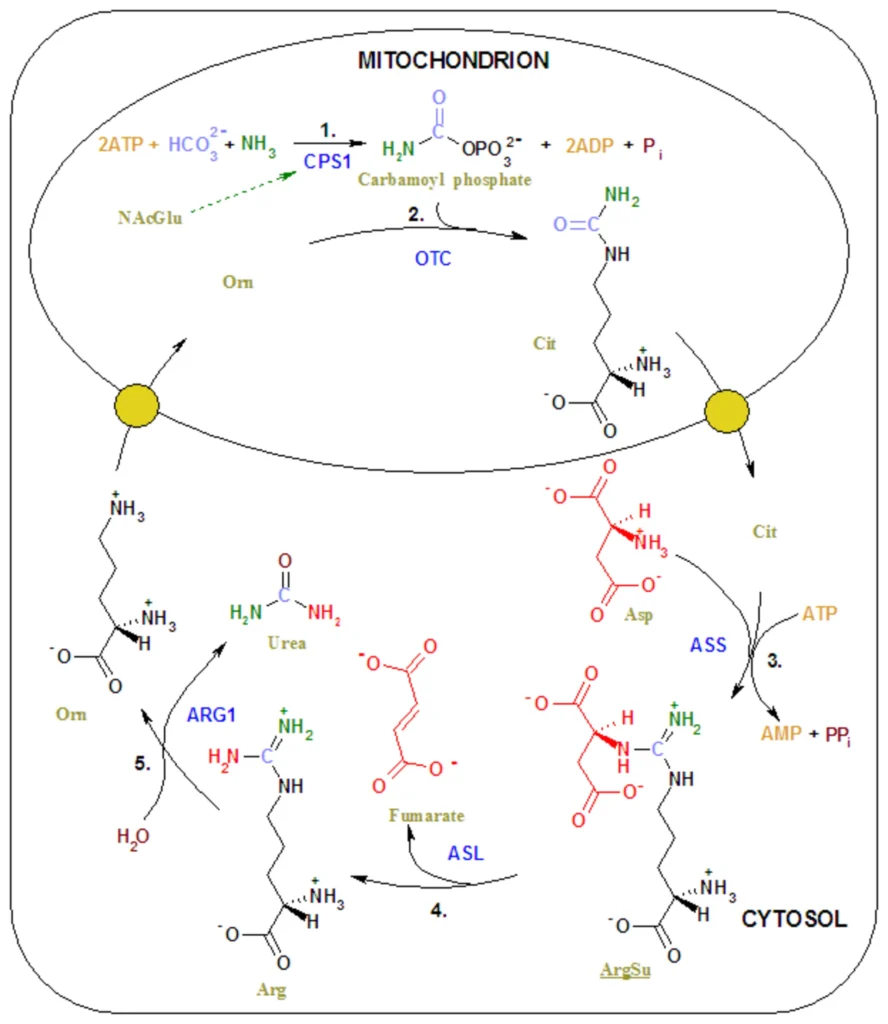
Occurs inside Liver cells (Mitochondria & Cytoplasm) to convert highly toxic ammonia into less toxic urea:
$\text{Amino Acid} \to \text{Glutamate} \to \text{NH}_3 + \text{CO}_2 + 2\text{ATP} \xrightarrow{\text{Carbamoyl Phosphate Synthetase}} \text{Carbamoyl Phosphate} + \text{Ornithine} \to \text{Citrulline} \xrightarrow{\text{Aspartate}} \text{Argininosuccinate} \to \text{Arginine} \xrightarrow{\text{Arginase}} \mathbf{\text{Urea}} + \text{Ornithine}$
B. Structure of Nephron
Structural and functional unit of kidney ($\approx 1\text{ million}$ per kidney). Each nephron consists of two parts:
- 1. Glomerulus: Tuft of capillaries formed by Afferent Arteriole (branch of renal artery) and drained by Efferent Arteriole.
- 2. Renal Tubule: Begins with cup-like Bowman’s Capsule (Glomerulus + Bowman’s Capsule = Malpighian Body / Renal Corpuscle).• Followed by Proximal Convoluted Tubule (PCT), Loop of Henle (Descending & Ascending limbs), and Distal Convoluted Tubule (DCT).
• DCT opens into Collecting Duct (Not a part of single nephron; collects from multiple nephrons).
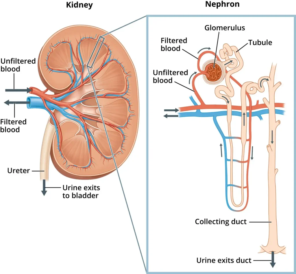
C. Types of Nephrons
| Feature | Cortical Nephrons (85%) | Juxtamedullary Nephrons (15%) |
|---|---|---|
| Loop of Henle (LOH) | Short LOH; extends only slightly into medulla. | Long LOH; runs deep into inner medulla. |
| Vasa Recta | Absent or highly reduced. | Well-developed capillary network surrounding LOH. |
| Function | Maintains normal urine formation. | Concentrates urine during water shortage / stress. |
3. Urine Formation & Glomerular Filtration Mechanics
A. Bowman’s Capsule & Filtration Membrane
- Visceral Layer: Lined by specialized podocyte cells with foot projections forming Filtration Slits / Slit Pores (25 nm wide).
- Parietal Layer: Simple squamous epithelium.
- 3-Layered Filtration Barrier: Capillary Endothelium (Fenestrated) $+$ Basement Membrane $+$ Visceral Podocytes.
B. Net Filtration Pressure (NFP) Forces
- 1. Glomerular Hydrostatic Pressure (GHP): $+60\text{ mmHg}$ (Pushes fluid out; due to Diameter of Afferent Arteriole $>$ Efferent Arteriole).
- 2. Blood Colloidal Osmotic Pressure (BCOP): $-30\text{ mmHg}$ (Opposes filtration; due to plasma proteins like albumin).
- 3. Capsular Hydrostatic Pressure (CHP): $-20\text{ mmHg}$ (Opposes filtration; pressure of fluid inside Bowman’s capsule).
- $$\mathbf{\text{NFP} = \text{GHP} – (\text{BCOP} + \text{CHP}) = 60 – (30 + 20) = 10\text{ mmHg}}$$
C. GFR, Renal Blood Flow & Filtration Fraction
- Renal Blood Flow (RBF): Kidneys receive $\frac{1}{5}\text{th}$ of cardiac output $= 1100\text{–}1200\text{ mL/min}$.
- Renal Plasma Flow (RPF): Plasma volume in RBF $\approx 600\text{ mL/min}$.
- Glomerular Filtration Rate (GFR): Amount of filtrate formed by kidneys per minute $= \mathbf{125\text{ mL/min}}\ (180\text{ L/day})$.
- Filtration Fraction (FF): $\frac{\text{GFR}}{\text{RPF}} = \frac{125}{600} \approx 16\text{–}20\%$.
- Ultrafiltrate Nature: Deproteinized plasma (contains $\text{H}_2\text{O}$, glucose, amino acids, urea, uric acid, salts; no blood cells or proteins).
4. Tubular Reabsorption, Secretion & Segmental Functions
A. Threshold Substances
- High Threshold: Maximally reabsorbed (e.g., Glucose, Amino acids, $\text{H}_2\text{O}$, $\text{Na}^+$).• Renal Threshold for Glucose: $180\text{ mg/dL}$ of blood. Exceeding this causes glucosuria.
- Low Threshold: Partially reabsorbed (e.g., Urea, Uric acid).
- Non-Threshold: Not reabsorbed at all; completely excreted (e.g., Inulin, Creatinine, Hippuric acid).
B. Functions of Tubular Segments
- 1. Proximal Convoluted Tubule (PCT):• Lined by simple cuboidal brush-border epithelium with dense microvilli and abundant mitochondria.
• Reabsorbs 70–80% of total filtrate ($100\%$ Glucose & Amino acids via active transport; $\frac{2}{3}\text{rd}$ $\text{H}_2\text{O}$, $\text{Na}^+$, $\text{Cl}^-$, $\text{HCO}_3^-$ passively).
• Osmolality remains Isotonic to blood plasma ($300\text{ mOsmol/L}$).
• Selective secretion of $\text{H}^+$, $\text{NH}_4^+$, and $\text{K}^+$ to maintain pH and ionic balance.
- 2. Loop of Henle (LOH):• Descending Limb: Permeable to $\text{H}_2\text{O}$, impermeable to salts. Filtrate becomes concentrated $\to$ Hypertonic ($1200\text{ mOsmol/L}$) at hair-pin bend.
• Ascending Limb: Impermeable to $\text{H}_2\text{O}$, permeable to salts ($\text{Na}^+, \text{Cl}^-$). Filtrate gets diluted $\to$ Hypotonic ($200\text{–}300\text{ mOsmol/L}$) as it enters cortex.
- 3. DCT & Collecting Duct: Conditional reabsorption of $\text{Na}^+$ and $\text{H}_2\text{O}$ under hormonal influence (Aldosterone & ADH).
5. Counter-Current Mechanism & Hormonal Control
A. Counter-Current Multiplier & Exchanger Mechanism
Operated by the Loop of Henle and Vasa Recta in Juxtamedullary nephrons to maintain an increasing osmotic gradient in medullary interstitium ($300\text{ mOsmol/L}$ in cortex to $1200\text{ mOsmol/L}$ in inner medulla).
- Gradient established primarily by $\text{NaCl}$ (transported by ascending LOH and exchanged with descending Vasa Recta) and Urea (recycled from collecting duct into thin ascending LOH).
- Allows human kidneys to produce urine that is 4 times more concentrated ($1200\text{ mOsmol/L}$) than initial filtrate ($300\text{ mOsmol/L}$).
B. Hormonal Feedback Mechanisms
- 1. RAAS System (Renin-Angiotensin-Aldosterone System):• Triggered by drop in Blood Pressure / Blood Volume / GFR.
• Sensed by Juxtaglomerular (JG) Cells of JGA $\to$ Releases enzyme Renin into blood.
• $\text{Angiotensinogen (from Liver)} \xrightarrow{\text{Renin}} \text{Angiotensin I} \xrightarrow{\text{ACE (Lungs)}} \text{Angiotensin II}$.
• Angiotensin II Effects: Powerful vasoconstrictor (raises BP) $+$ Stimulates Adrenal Cortex to release Aldosterone (increases $\text{Na}^+$ and $\text{H}_2\text{O}$ reabsorption from DCT) $\to$ Restores GFR.
- 2. ADH / Vasopressin Control:• High blood osmolality / fluid loss triggers Hypothalamus osmoreceptors $\to$ Posterior Pituitary releases Anti-Diuretic Hormone (ADH).
• Facilitates water reabsorption through aquaporins in DCT and Collecting Duct $\to$ Prevents Diuresis.
- 3. ANF Factor (Check on RAAS):• Increase in blood volume/pressure causes Atria of Heart to release Atrial Natriuretic Factor (ANF).
• Causes Vasodilation and promotes $\text{Na}^+$ excretion (Natriuresis) $\to$ Decreases BP (acts as counter-check to RAAS).
6. Micturition, Urine Properties & Disorders
A. Micturition Reflex & Normal Urine Properties
- Micturition Reflex: Bladder filling stretches walls $\to$ Stretch receptors send signals to CNS $\to$ Motor signals induce contraction of Detrusor muscle and relaxation of urethral sphincters.
- Daily Excretion Volume: $1.0\text{ to } 1.5\text{ Litres}$ of urine per day ($25\text{–}30\text{ g}$ of Urea excreted daily).