
Body Fluids and Circulation
1. Blood Composition, RBCs & Leucocytes (WBCs)
Blood: Mesodermal origin, fibre-less fluid connective tissue. Slightly alkaline ($\text{pH} \approx 7.4$), heavier than water.
A. Plasma (55% Matrix) & Formed Elements (45%)
- Plasma Composition: Pale yellow / straw-coloured fluid part. $90\text{–}92\%$ Water, $6\text{–}8\%$ Plasma Proteins.• Albumin: Most abundant; holds colloid water; maintains Blood Colloidal Osmotic Pressure (BCOP) and blood volume.
• Globulin: $\alpha, \beta$ transport lipids; $\gamma$-globulins act as Antibodies for body defence/immunity.
• Fibrinogen: Major clotting protein; prevents excess blood loss.
- Serum: Plasma without clotting factors ($\text{Serum} = \text{Plasma} – \text{Fibrinogen}$).
B. RBCs (Erythrocytes)
- Count: $5\text{–}5.5\text{ million/mm}^3$ of blood. Formed via Erythropoiesis in red bone marrow (induced by Erythropoietin hormone from kidney; requires Vit $B_{12}$, Folic acid, and Iron).
- Structure & Lifespan: Biconcave, enucleated at maturity (lacks nucleus, mitochondria, and organelles to carry more Haemoglobin). Lifespan is 120 days. Survives anaerobically. Graveyard of RBC is the Spleen.
- Haemoglobin ($Hb$): $12\text{–}16\text{ g / 100 mL}$ of blood.• Deficiency: Erythrocytopenia / Anaemia.
• Excess: Polycythemia (blood becomes viscous, reducing $O_2$ flow).
C. WBCs (Leucocytes) Classification
Normal Count: $6000\text{–}8000\text{ / mm}^3$. Low count = Leucopenia; High count = Leucocytosis (infection site); Extremely high = Leukaemia (blood cancer). Formation process = Leucopoiesis.
| Category | Cell Type | % Share & Key Features |
|---|---|---|
| Granulocytes (Granular cytoplasm) | Neutrophil | $60\text{–}65\%$ (Most abundant). Multilobed nucleus; PMNL (Polymorphonuclear leucocyte). Stained by both acidic/basic dyes. First line of defence via Phagocytosis. |
| Basophil | $0.5\text{–}1\%$ (Least abundant). S-shaped nucleus; stained by basic dyes. Secretes Heparin (anticoagulant), Histamine, and Serotonin during inflammatory reactions. | |
| Eosinophil | $2\text{–}3\%$. Bilobed nucleus; stained by acidic dyes (Eosin). Resists infections, involved in allergic reactions, kills helminthic larvae. | |
| Agranulocytes (Agranular cytoplasm) | Monocyte | $6\text{–}8\%$. Largest WBC with kidney/bean-shaped nucleus. Phagocytic in nature. |
| Lymphocyte | $20\text{–}25\%$. Large round nucleus. B-lymphocytes (humoral immunity via antibody production) & T-lymphocytes (cell-mediated immunity). |
2. Platelets, Blood Grouping & Coagulation Cascade
A. Platelets (Thrombocytes)
- Non-cellular cell fragments produced from Megakaryocytes in bone marrow.
- Count: $1.5\text{–}3.5\text{ lakh/mm}^3$. Lifespan: $7\text{–}8\text{ days}$. Low count = Thrombocytopenia (leads to bleeding disorders).
- Function: Secretes platelet factors involved in blood clotting.
B. ABO & Rh Blood Grouping Systems
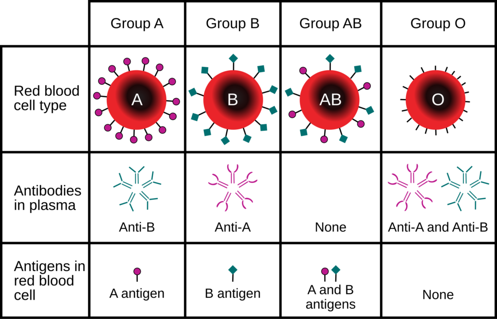
| Blood Group | Antigen on RBC | Antibody in Plasma | Compatible Donors |
|---|---|---|---|
| A | A | Anti-B | A, O |
| B | B | Anti-A | B, O |
| AB | A, B | Nil | AB, A, B, O (Universal Recipient) |
| O | Nil | Anti-A, Anti-B | O (Universal Donor) |
C. Blood Clotting Mechanism (Cascade Process)
Involves 13 clotting factors operating via Extrinsic and Intrinsic pathways:
Injury / Platelets $\to$ Release Thromboplastin (Factor III) $\to$ Forms Thrombokinase complex $\xrightarrow{Ca^{2+}}$ Inactive Prothrombin (Factor II) $\to$ Active Thrombin $\to$ Soluble Fibrinogen (Factor I) $\to$ Insoluble Fibrin mesh (Clot) (Stabilized by Factor XIII).
Vitamin K and $Ca^{2+}$ (Factor IV) are mandatory for clotting.
3. Lymph, Circulatory Pathways & Vertebrate Hearts
A. Lymph (Tissue Fluid)
- Colourless fluid containing specialized lymphocytes. Acts as “Middleman of the body” for exchange of nutrients, gases, and hormones between blood and cells.
- Fats are absorbed through Lacteals in intestinal villi via the lymphatic system.
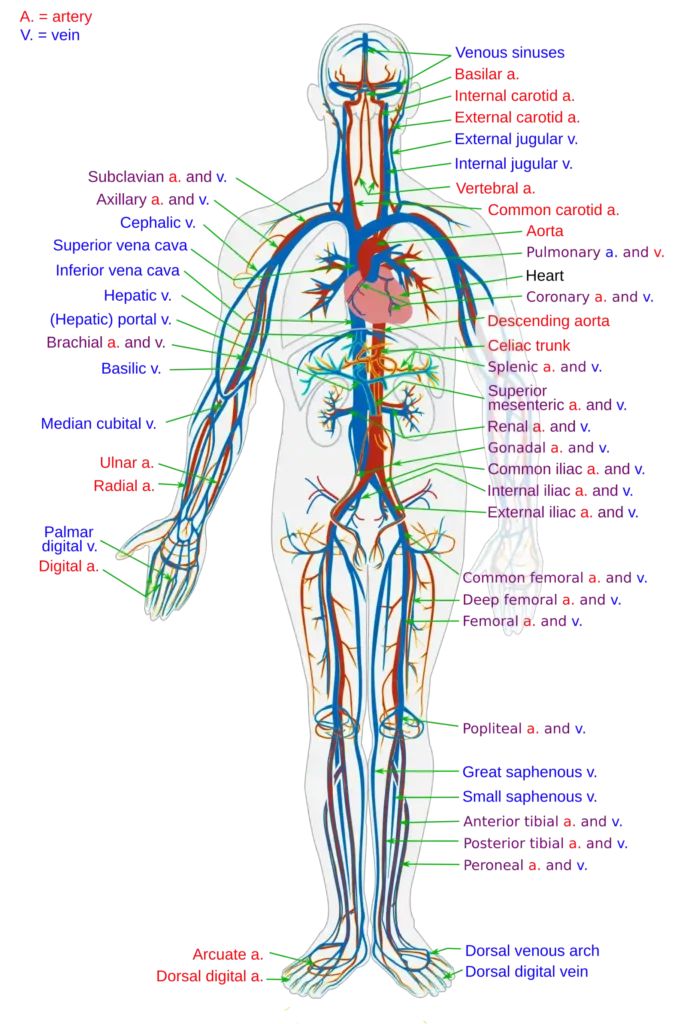
B. Open vs. Closed Circulatory Systems
| Feature | Open Circulatory System | Closed Circulatory System |
|---|---|---|
| Blood Flow | Blood fills open body sinuses (Haemocoel). | Blood flows within closed network of vessels. |
| Pressure & Speed | Low pressure, slow velocity. | High pressure, high velocity (rapid exchange). |
| Examples | Arthropoda, Mollusca (except Cephalopods), Echinodermata. | Annelids (Earthworm – 1st closed system), Cephalopod Molluscs, Vertebrates. |
C. Evolution of Vertebrate Heart Structure
- Fishes (2-Chambered): 1 Atrium + 1 Ventricle (plus accessory Sinus Venosus & Conus Arteriosus). Venous heart pumping deoxygenated blood only $\to$ Single Circulation.
- Amphibians & Reptiles (3-Chambered): 2 Atria + 1 Ventricle. Mixed blood in ventricle $\to$ Incomplete Double Circulation. (Exception: Crocodiles have 4-chambered heart).
- Birds & Mammals (4-Chambered): 2 Atria + 2 Ventricles. Completely separated oxygenated and deoxygenated blood $\to$ Double Circulation.
4. Human Heart Anatomy & Cardiac Cycle
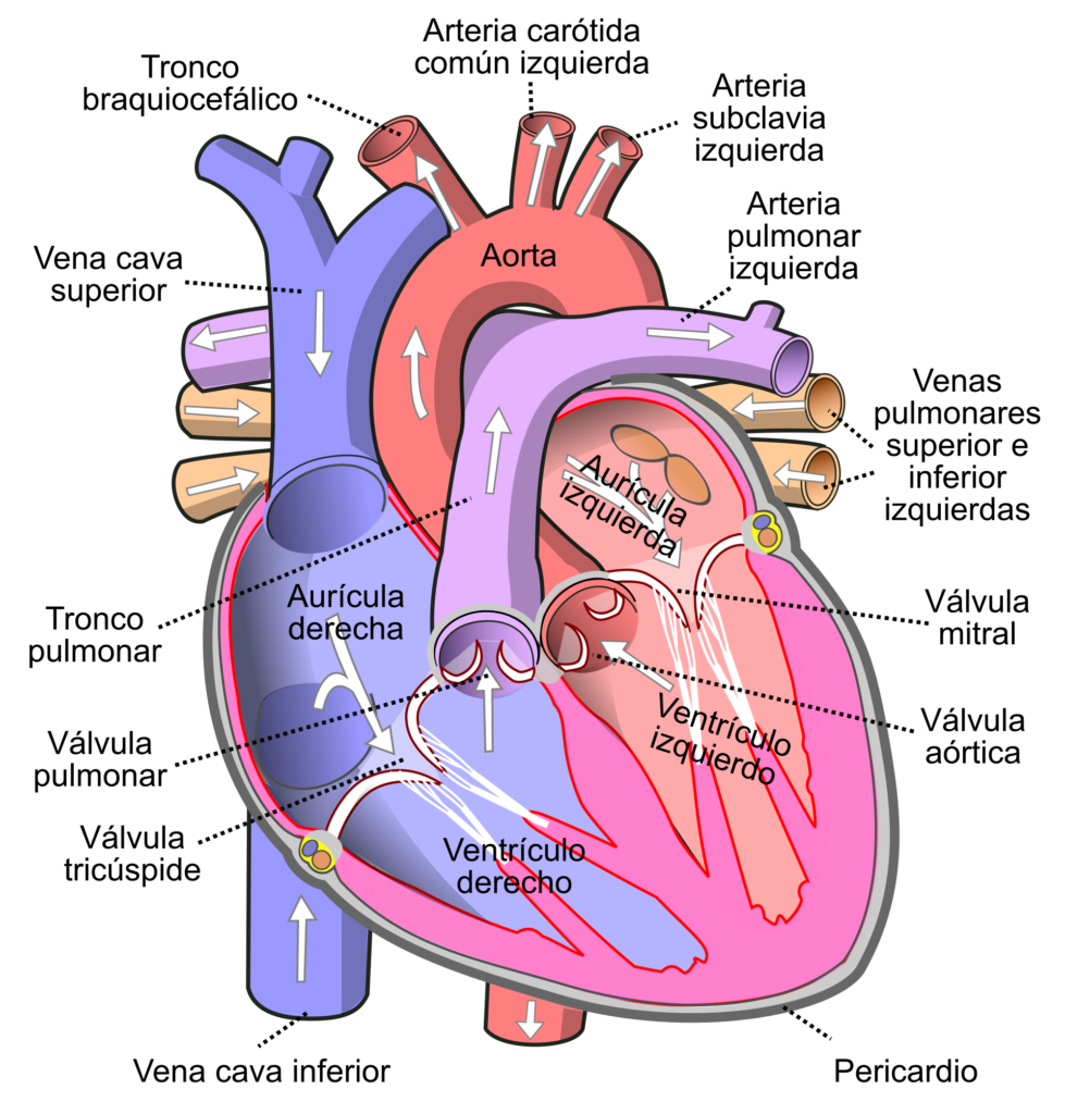
A. Anatomy & Internal Chambers
- Location & Size: Clenched fist-sized conical organ located in thoracic cavity inside mediastinum, slightly tilted left. Protected by double-walled Pericardium (outer fibrous, inner serous with pericardial fluid).
- Heart Wall Histology (3 Layers): Outer Epicardium, Middle thick muscular Myocardium (cardiac muscle), Inner Endocardium (simple squamous).
- Chambers & Valves:• Right Atrium: Receives deoxygenated blood via Superior Vena Cava (upper body), Inferior Vena Cava (lower body – guarded by Eustachian valve), and Coronary Sinus (heart wall – guarded by Thebesian valve).
• Right Ventricle: Separated from RA by Tricuspid Valve (3 cusps). Pumps blood to lungs via Pulmonary Artery (guarded by Semilunar valve).
• Left Atrium: Receives oxygenated blood via 4 Pulmonary Veins (2 from each lung).
• Left Ventricle: Thickest muscular wall. Separated from LA by Bicuspid / Mitral Valve (2 cusps). Pumps blood to body via Aorta (guarded by Semilunar valve).
• Chordae Tendinae: White fibrous cords connecting valve cusps to Papillary muscles of ventricular wall to prevent backflow during systole.
B. Cardiac Cycle Phases (Duration: 0.8 Seconds at 72 bpm)
- 1. Atrial Systole (0.1 sec): Both atria contract, pushing remaining 30% blood into ventricles. AV valves open.
- 2. Ventricular Systole (0.3 sec): Ventricles contract.• Pressure rises $\to$ AV valves close $\to$ Produces First Heart Sound (LUB) (longer duration, lower pitch).
• Pressure exceeds aorta/pulmonary artery $\to$ Semilunar valves open $\to$ Blood ejected.
- 3. Joint Diastole (0.4 sec): All 4 chambers relax.• Ventricular pressure falls $\to$ Semilunar valves close $\to$ Produces Second Heart Sound (DUB) (shorter duration, higher pitch).
5. Nodal Tissue, Electrocardiogram (ECG) & Blood Pressure
A. Conducting System of Heart (Myogenic Heart)
Human heart generates auto-excitable action potentials through specialized nodal cardiac musculature:
SAN (Sino-Atrial Node – Pacemaker, 70–75/min) $\to$ AVN (Atrio-Ventricular Node – Pace-setter) $\to$ Bundle of His $\to$ Purkinje Fibres (Ventricular walls)
B. Electrocardiogram (ECG) Waves
Graphical representation of the electrical activity of the heart during a cardiac cycle (Discovered by Einthoven – Nobel Prize):
- P Wave: Represents electrical excitation / Atrial Depolarization (leads to atrial contraction).
- QRS Complex: Represents Ventricular Depolarization (initiates ventricular contraction). Counting QRS complexes in a given time period gives heart rate/pulse rate.
- T Wave: Represents return of ventricles to relaxed state / Ventricular Repolarization. End of T wave marks end of systole.
- Clinical Deviations: $ST$ segment elevation indicates Myocardial Infarction (Heart Attack); $ST$ depression indicates Ischemia.
C. Hemodynamic Volumes & Blood Pressure
- Stroke Volume (SV): Volume of blood pumped out by each ventricle per beat ($\approx 70\text{ mL}$).$$\text{SV} = \text{End Diastolic Volume (120 mL)} – \text{End Systolic Volume (50 mL)} = 70\text{ mL}$$
- Cardiac Output (CO): Volume of blood pumped by each ventricle per minute.$$\text{CO} = \text{Stroke Volume} \times \text{Heart Rate} = 70\text{ mL} \times 72\text{ bpm} \approx 5000\text{ mL (5 Litres/min)}$$
(Can increase up to 30 L/min in athletes). - Blood Pressure (BP): Measured using Sphygmomanometer. Normal BP = $120 / 80\text{ mmHg}$ ($120\text{ mmHg}$ Systolic / $80\text{ mmHg}$ Diastolic).• Pulse Pressure: $\text{Systolic BP} – \text{Diastolic BP} = 120 – 80 = 40\text{ mmHg}$.
6. Regulation, Portal Systems, Blood Vessels & Disorders
A. Regulation of Cardiac Activity
- Autonomic Nervous System (ANS): Neural centre in Medulla Oblongata.• Sympathetic Nervous System: Secretes Adrenaline/Noradrenaline $\to$ Increases heart rate, ventricular contraction force, and Cardiac Output.
• Parasympathetic Nervous System: Secretes Acetylcholine $\to$ Decreases heart rate and Cardiac Output to normal.
- Hormonal Control: Adrenal medullary hormones (Adrenaline/Noradrenaline) increase cardiac output during stress/emergency.
B. Portal Circulation Systems
- Hepatic Portal System: Deoxygenated blood from intestine/digestive tract is carried to the liver via Hepatic Portal Vein before reaching systemic circulation to process nutrients and toxins.
- Hypophyseal Portal System: Carries blood from Hypothalamus to Anterior Pituitary gland.
C. Blood Vessels Structure (Artery vs. Vein)
| Feature | Artery | Vein |
|---|---|---|
| Direction & Blood | Carries oxygenated blood away from heart (except Pulmonary Artery). | Carries deoxygenated blood toward heart (except Pulmonary Vein). |
| Wall Layers | Thick Tunica Media (smooth muscle & elastic fibers); narrow lumen. | Thin Tunica Media, prominent Tunica Externa; wide lumen. |
| Valves & Pressure | No valves; high pressure, jerky flow. Deep seated. | Valves present (prevents backflow); low pressure. Superficial. |
D. Cardiovascular Disorders
- 1. Hypertension (High BP): Sustained BP $> 140/90\text{ mmHg}$. Damages vital organs like heart, kidneys, and brain.
- 2. Coronary Artery Disease (CAD) / Atherosclerosis: Deposition of calcium, fat, cholesterol, and fibrous tissue in coronary arteries, narrowing lumen and reducing blood supply to heart muscle.
- 3. Angina Pectoris: Acute chest pain when insufficient oxygen reaches heart muscles.
- 4. Heart Attack (Myocardial Infarction): Sudden damage to heart muscle due to inadequate blood supply (ischemia).
- 5. Heart Failure: State when heart fails to pump blood effectively enough to meet body needs (often called congestive heart failure due to lung congestion).
- 6. Cardiac Arrest: Complete stoppage of heart beat.