
1. Plant Tissues: Meristematic & Simple Permanent Tissues
A. Classification of Plant Tissues
A group of cells having common origin and usually performing a common function (structures may be similar or different).
| Feature | Meristematic Tissue | Permanent Tissue |
|---|---|---|
| Cell Division Capacity | Actively dividing, small, living, compactly arranged cells with thin cellulosic walls. | Cells lose division capacity (temporarily or permanently) and differentiate to perform specific functions. |
| Cellular Features | Prominent central nucleus, dense cytoplasm, vacuoles absent or tiny, no $2^\circ$ wall. | Living or dead; mature shapes with specialized secondary wall thickenings. |
| Classification Types | • By Origin: Primary (SAM, RAM) vs. Secondary (Cork/Interfascicular Cambium). • By Position: Apical, Intercalary, Lateral (Vascular Cambium). | • Simple: Parenchyma, Collenchyma, Sclerenchyma. • Complex: Xylem, Phloem. |
B. Simple Permanent Tissues Comparison
- 1. Parenchyma: Living, universal, fundamental packaging tissue. Cells are isodiametric (spherical/oval/polygonal) with thin cellulosic primary walls and intercellular spaces (ICS).• Chlorenchyma: Contains chloroplasts; performs photosynthesis (e.g., Leaf Mesophyll).
• Aerenchyma: Large air cavities providing buoyancy to aquatic plants (Hydrophytes).
• Idioblasts: Special storage parenchyma storing gums, resins, tannins.
• Prosenchyma: Elongated parenchyma in Xylem and Phloem providing minor mechanical support.
- 2. Collenchyma: Living mechanical tissue present as a homogeneous layer or patches below epidermis (Hypodermis) in young Dicot stems and leaf petioles. ABSOLUTELY ABSENT IN MONOCOTS AND ROOTS!• Cells compactly arranged with localized Pectin, Cellulose, and Hemicellulose corner thickenings. Intercellular spaces absent. Provides mechanical strength with flexibility (elasticity).
- 3. Sclerenchyma: Dead mechanical tissue at maturity lacking protoplasm. Cells have thick, highly Lignified walls with narrow lumen.• Fibres: Long, narrow, elongated cells with pointed/tapering ends (e.g., Xylem fibres, Phloem fibres).
• Sclereids (Stone Cells): Spherical/oval cells with extremely narrow lumen/cavity (e.g., Fruit wall of nuts, pulp of Guava, Pear, Sapota, seed coat of legumes, tea leaves).
2. Complex Permanent Tissues: Xylem & Phloem Mechanics
A. Xylem Components (Water & Mineral Transport)
- 1. Tracheids: Dead, elongated tube-like cells with thick lignified walls and tapering ends. Present in Pteridophytes, Gymnosperms, and Angiosperms. Main water conducting element in Gymnosperms.
- 2. Vessels: Long cylindrical tube structures made of many vessel elements with perforated end walls (continuous pipeline). Dead and lignified. Characteristic feature of Angiosperms (Absent in Gymnosperms except Gnetum, Ephedra, Welwitschia).
- 3. Xylem Fibres: Dead sclerenchymatous fibres with highly thickened walls and obliterated central lumens. Provide mechanical support.
- 4. Xylem Parenchyma: ONLY LIVING COMPONENT of Xylem. Thin-walled parenchymatous cells storing food (starch/fat) and tannins. Ray parenchyma cells perform radial conduction of water.
• Endarch Condition (Stems): Protoxylem lies toward center (pith) and Metaxylem lies toward periphery.
B. Phloem Components (Food & Hormone Transport)
- 1. Sieve Tube Elements: Living, elongated cells arranged end-to-end with perforated end walls forming Sieve Plates. A mature sieve element possesses peripheral cytoplasm and a large vacuole, but LACKS A NUCLEUS! Functions controlled by nucleus of companion cell.
- 2. Companion Cells: Specialized parenchymatous cells connected to sieve tube elements via pit fields. Maintain pressure gradient in sieve tubes. Absent in Gymnosperms & Pteridophytes (replaced by Albuminous Cells / Sieve Cells).
- 3. Phloem Parenchyma: Living, elongated parenchymatous cells with dense cytoplasm. Store resins, latex, mucilage. ABSENT IN MOST MONOCOTS!
- 4. Phloem Fibres (Bast Fibres): ONLY DEAD COMPONENT of Phloem. Sclerenchymatous fibres providing mechanical strength. Commercial bast fibres obtained from Jute, Flax, Hemp. Absent in primary phloem, present in secondary phloem.
3. Tissue Systems, Stomatal Types & Vascular Bundles
A. Epidermal Tissue System & Appendages
- Epidermis: Outermost single parenchymatous layer coated with waxy Cuticle (prevents transpirational water loss; cuticle absent in roots!).
- Root Hairs: Unicellular, unbranched exogenous extensions of epidermal cells (Epiblema) specialized for water absorption.
- Trichomes: Multicellular or unicellular, branched or unbranched exogenous shoot epidermal hairs; reduce water loss and may be secretory.
- Stomatal Apparatus: Consists of Stomatal Pore $+$ 2 Guard Cells $+$ Subsidiary Cells.• Dicot Guard Cells: Kidney / Bean-shaped with thick inner cellulosic walls and thin elastic outer walls.
• Monocot Guard Cells: Dumb-bell shaped (e.g., Grasses).
B. Vascular Bundle Types
| Vascular Arrangement | Structural Feature | Occurrence / Examples |
|---|---|---|
| 1. Radial Bundles | Xylem and Phloem arranged in separate alternating radii. | All Roots (Dicot & Monocot). |
| 2. Conjoint Collateral Open | Xylem (inside) and Phloem (outside) on same radius with Vascular Cambium strip present between them. Capable of $2^\circ$ growth. | Dicot Stems & Gymnosperm stems. |
| 3. Conjoint Collateral Closed | Xylem and Phloem on same radius LACKING cambium. Incapable of $2^\circ$ growth. | Monocot Stems & Leaves. |
| 4. Bicollateral Bundles | Phloem-Cambium-Xylem-Cambium-Phloem sandwich. Always open. | Cucurbitaceae (Gourds, Pumpkin). |
C. Ground Tissue System & Stele Definition
- All tissues except epidermal and vascular systems form ground tissue. Comprises Hypodermis, Cortex, Endodermis, Pericycle, Pith, and Medullary Rays.
- Stele: All tissues inner to endodermis, i.e., Pericycle $+$ Vascular Bundles $+$ Pith $+$ Medullary Rays.
4. Comparative Internal Anatomy: Dicot vs. Monocot Root & Dicot Stem
A. Dicot Root vs. Monocot Root Anatomy
| Anatomical Feature | Dicot Root (e.g., Sunflower Root) | Monocot Root (e.g., Maize Root) |
|---|---|---|
| Xylem Bundle Count | Diarch to Hexarch (2 to 4–6 patches). Exarch. | Polyarch ($> 6$ xylem patches, usually many). Exarch. |
| Pith Region | Small, poorly developed or completely absent. | Large, well-developed parenchymatous pith. |
| Secondary Growth | Present (Vascular cambium formed from conjunctive tissue & pericycle). | ABSOLUTELY ABSENT. |
| Endodermis Features | Barrel-shaped cells with Casparian Strips (suberin deposition on radial and tangential walls acting as water checkpoint). Passage cells opposite protoxylem. | |
B. Anatomy of Dicot Stem (e.g., Sunflower Stem)
- Epidermis: Single layer with cuticle, stomata, and multicellular trichomes.
- Cortex (3 Sub-layers):1. Hypodermis: Few layers of Collenchymatous cells providing mechanical strength to young stem.
2. Cortical Layers: Thin-walled parenchymatous cells with intercellular spaces.
3. Endodermis (Starch Sheath): Innermost layer rich in starch grains.
- Pericycle: Sclerencymatous semi-lunar patches above phloem bundles.
- Vascular Bundles: Arranged in a Ring (Eustele). Conjoint, Collateral, Open with Endarch protoxylem. Medullary rays present between bundles. Pith large and parenchymatous.
5. Monocot Stem Anatomy & Leaf Histology
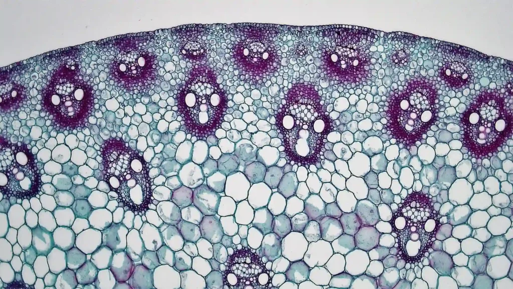
A. Anatomy of Monocot Stem (e.g., Maize Stem)
- Epidermis: Coated with cuticle; trichomes absent.
- Hypodermis: Sclerenchymatous layer (differs from collenchymatous dicot hypodermis!).
- Ground Tissue: Undifferentiated large parenchymatous mass; cortex, endodermis, pericycle, and pith are NOT differentiated.
- Vascular Bundles:• Numerous Scattered vascular bundles (Atactostele) throughout ground tissue. Peripheral bundles smaller and closer; central ones larger.
• Conjoint, Collateral, Closed (No cambium $\to$ No secondary growth). Each bundle surrounded by a Sclerenchymatous Bundle Sheath.
• Lysigenous Water Cavities (Protoxylem Cavity): Present within vascular bundles due to breakdown of inner protoxylem vessels.
• Phloem Parenchyma is ABSENT!
B. Dorsiventral (Dicot) vs. Isobilateral (Monocot) Leaf Summary
| Feature | Dorsiventral Leaf (Dicot) | Isobilateral Leaf (Monocot) |
|---|---|---|
| Stomatal Distribution | Hypostomatic (More stomata on lower abaxial surface). | Amphistomatic (Equal stomata on adaxial and abaxial surfaces). |
| Mesophyll Differentiation | Differentiated into upper Palisade Parenchyma (columnar, vertical) and lower Spongy Parenchyma (loosely packed with air cavities). | Undifferentiated mesophyll (all spongy/isodiametric cells with small air spaces). |
| Special Cellular Features | Reticulate venation; vascular bundles vary in size according to vein diameter. | Parallel venation. Possesses large, empty, colourless Bulliform Cells on upper epidermis (cause leaf rolling during water stress to minimize transpiration). |