🧬 Molecular Basis of Inheritance – The Blueprint of Life
1. Discovery of Nucleic Acids & DNA Double Helix
A. Historical Context & Basic Definitions
- Friedrich Miescher (1869): First extracted acidic substance from nucleus of pus cells/owl cells and named it Nuclein.
- Altmann: Proved acidic nature of nuclein and renamed it Nucleic Acid.
• DNA (Deoxyribonucleic Acid): Genetic material in most organisms; most abundant genetic material.
• RNA (Ribonucleic Acid): Genetic material in some viruses (TMV, HIV, QB Bacteriophage); acts as messenger, adapter, structural, and catalytic molecule.
B. DNA Length across Organisms
| Organism | Type of DNA | Number of Nucleotides / Base Pairs |
|---|---|---|
| Human (Haploid $n$) | ds Linear DNA | $3.3 \times 10^9\text{ bp}$ ($6.6 \times 10^9\text{ bp}$ in diploid $2n$) |
| Escherichia coli | ds Circular DNA | $4.6 \times 10^6\text{ bp}$ |
| Bacteriophage $\lambda$ | ds Linear DNA | $48,502\text{ bp}$ |
| $\phi \times 174$ Bacteriophage | ss Circular DNA | $5,386\text{ nucleotides/bases}$ |
C. Chemical Composition of Nucleotides
- $$\mathbf{\text{Nucleoside} = \text{Pentose Sugar} + \text{Nitrogenous Base}}$$
- $$\mathbf{\text{Nucleotide} = \text{Pentose Sugar} + \text{Nitrogenous Base} + \text{Phosphate Group}}$$
- Nitrogenous Bases:
• Purines (9-membered double ring; $N$ at 1,3,7,9): Adenine ($A$) and Guanine ($G$).
• Pyrimidines (6-membered single ring; $N$ at 1,3): Cytosine ($C$), Thymine ($T$ – in DNA), Uracil ($U$ – in RNA).
- Chemical Bonds:
• N-Glycosidic Linkage: Connects $C_1’$ of sugar with $N_9$ of Purine or $N_1$ of Pyrimidine.
• Phosphoester Linkage: Connects phosphate group to $C_5’$ OH of nucleoside.
• $3’\text{–}5’$ Phosphodiester Bond: Connects adjacent nucleotides in a polynucleotide chain.
D. Chargaff’s Rules (Only for dsDNA)
- Molar concentration of Adenine $=$ Thymine ($[A] = [T]$) and Guanine $=$ Cytosine ($[G] = [C]$).
- Total Purines $=$ Total Pyrimidines $\implies \frac{A + G}{T + C} = 1$ (Constant for all species).
- Base Ratio ($\frac{A + T}{G + C}$): Specific for a species. $>1$ in eukaryotes (Human $= 1.52$), $<1$ in prokaryotes ($E. coli = 0.93$).
2. DNA Double Helix Features & Packaging (Nucleosome)
A. Watson & Crick Model of DNA (1953)
Based on X-ray diffraction data of Maurice Wilkins & Rosalind Franklin and Chargaff’s equivalence rules:
- Two polynucleotide chains run in antiparallel polarity ($5′ \to 3’$ and $3′ \to 5’$). Backbone formed by sugar-phosphate, bases project inside.
- Right-handed helical coiling. Diameter $= 20\text{ \AA}\ (2\text{ nm})$.
- Pitch of Helix: $3.4\text{ nm}\ (34\text{ \AA})$ per turn; contains $10\text{ base pairs}$. Distance between adjacent base pairs $= 0.34\text{ nm}\ (3.4\text{ \AA})$.
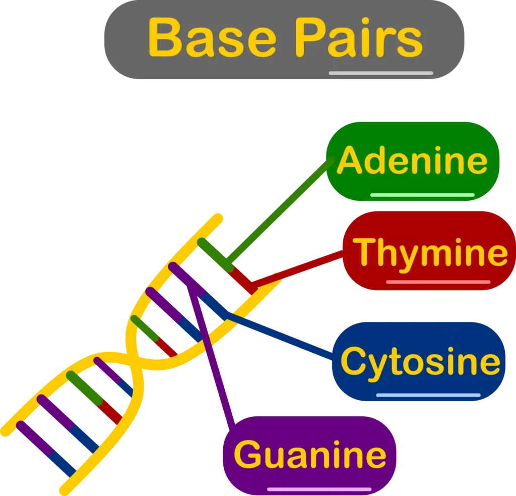
- Hydrogen Bonding: $A = T$ (2 Hydrogen bonds), $G \equiv C$ (3 Hydrogen bonds). Base stacking provides thermodynamic stability.
B. Formulas for Bond Calculations
| DNA Type | Phosphodiester Bonds | Glycosidic Bonds |
|---|---|---|
| Single-Stranded Linear ($n = \text{nucleotides}$) | $n – 1$ | $n$ |
| Double-Stranded Linear ($N = \text{total nucleotides}$) | $N – 2$ | $N$ |
| Single-Stranded Circular | $n$ | $n$ |
| Double-Stranded Circular | $N$ | $N$ |
C. Packaging of DNA (Nucleosome Model)
- Length of Human DNA: $6.6 \times 10^9\text{ bp} \times 0.34 \times 10^{-9}\text{ m/bp} = \mathbf{2.2\text{ metres}}$.
- Prokaryotes ($E. coli$ DNA length $= 1.36\text{ mm}$): No nucleus, but DNA is held in nucleoid by positively charged non-histone basic proteins.
- Eukaryotic Packaging: Negatively charged acidic DNA wrapped around positively charged basic Histone Octamer (rich in basic amino acids Lysine and Arginine).
• Histone Octamer Core: Contains 2 copies each of $H_2A, H_2B, H_3, H_4$ (8 proteins).
• Nucleosome Unit: Octamer core $+$ $H_1$ linker protein $+$ $\mathbf{200\text{ bp}}$ wrapped DNA. Under electron microscope appears as “Beads-on-string” structure.
3. Chromatin Types & Search for Genetic Material
A. Euchromatin vs. Heterochromatin
| Feature | Euchromatin | Heterochromatin |
|---|---|---|
| Staining & Density | Lightly stained; loosely packed region. | Densely stained; highly supercoiled region. |
| Transcriptional Activity | Transcriptionally Active. | Transcriptionally Inactive. |
B. Griffith’s Transformation Experiment (1928)
- Worked on Streptococcus pneumoniae using S-strain (Smooth capsule, Virulent) and R-strain (Rough, Non-virulent):
• $\text{S-strain} \to \text{Injected into mice} \to \text{Mice Die}$.
• $\text{R-strain} \to \text{Injected into mice} \to \text{Mice Live}$.
• $\text{Heat-killed S-strain} \to \text{Injected into mice} \to \text{Mice Live}$.
• $\text{Heat-killed S-strain} + \text{R-strain} \to \text{Injected into mice} \to \mathbf{\text{Mice Die}}$ (Living S-strain recovered).
- Conclusion: Non-virulent R-strain transformed into virulent S-strain by absorbing a ‘Transforming Principle’.
C. Biochemical Characterisation (Avery, MacLeod, McCarty – 1944)
- Purified proteins, RNA, and DNA from heat-killed S-cells to test transformation:
• $\text{Proteases} + \text{RNase treatment} \implies \text{Transformation still occurred}$.
• $\mathbf{\text{DNase treatment}} \implies \mathbf{\text{Transformation inhibited}}$.
- Proved DNA is the transforming genetic material.
D. Hershey & Chase Transduction Experiment (1952)
Unequivocal proof that DNA is genetic material using Bacteriophage $T_2$ and radioactive isotopes:
- Radioactive $\mathbf{^{32}P}$ (Labels DNA): Radioactivity detected in bacterial pellet (cell).
- Radioactive $\mathbf{^{35}S}$ (Labels Protein Coat): Radioactivity detected only in supernatant (viral coats).
- Steps: Infection $\to$ Blending (detached coat) $\to$ Centrifugation. Proved viral DNA enters host cell.
4. DNA vs. RNA & Central Dogma
A. Properties of Genetic Material & DNA vs RNA Comparison
Essential criteria for genetic material: Replication capability, Chemical stability, Scope for slow mutations, and Expression of Mendelian traits.
| Feature | DNA | RNA |
|---|---|---|
| Sugar & Base | Deoxyribose sugar; Bases: $A, T, C, G$. | Ribose sugar; Bases: $A, U, C, G$. |
| Chemical Stability | Highly stable (Lacks $2’\text{-OH}$ group; contains 5-Methyl Uracil / Thymine). Preferred for storage of genetic information. | Unstable & Reactive ($2’\text{-OH}$ makes it catalytic and easily degradable). Preferred for transmission of genetic information. |
| Mutation Rate | Mutates at a slower, controlled rate. | Mutates faster (viruses with RNA genome short-lived & evolve rapidly). |
B. Central Dogma & Reverse Transcription
Proposed by Francis Crick. Describes unidirectional flow of genetic information:
$$\mathbf{\text{DNA}} \xrightarrow{\text{Replication}} \text{DNA} \xrightarrow{\text{Transcription}} \mathbf{\text{RNA}} \xrightarrow{\text{Translation}} \mathbf{\text{Protein}}$$
- Reverse Transcription (Teminism): Discovered by Temin & Baltimore in Rous Sarcoma Virus (RSV). Genetic information flows backwards from $\text{RNA} \to \text{DNA}$ via Reverse Transcriptase enzyme.
5. DNA Replication & Semi-Conservative Proof
A. Semi-Conservative Replication Mechanism
- Proposed by Watson & Crick. Each daughter DNA molecule retains one parental strand and synthesizes one new complementary strand.
- Occurs during S-phase of cell cycle in nucleus (Eukaryotes) or cytoplasm (Prokaryotes).
B. Experimental Proofs
- Meselson & Stahl Experiment (1958):
• Grew $E. coli$ in heavy nitrogen $\text{N}^{15}$ medium, then transferred to light $\text{N}^{14}$ medium.
• Centrifuged in $\text{CsCl}$ density gradient.
• Generation I (20 mins): $100\%$ Hybrid DNA ($\text{N}^{15}\text{-N}^{14}$).
• Generation II (40 mins): $50\%$ Hybrid ($\text{N}^{15}\text{-N}^{14}$) $+$ $50\%$ Light ($\text{N}^{14}\text{-N}^{14}$).
- Taylor et al. Experiment (1958): Proved semi-conservative replication at chromosomal level in plant Vicia faba (faba beans) using Radioactive Thymidine ($^3\text{H-Thymidine}$).
6. Enzymes of Replication & Y-Shaped Replication Fork
A. Enzymes Involved in Replication
- 1. Helicase: Unwinds double helix by breaking hydrogen bonds (ATP dependent).
- 2. Topoisomerase / DNA Gyrase: Relieves torsional strain and supercoiling ahead of unwinding fork.
- 3. SSBP (Single-Stranded Binding Proteins): Prevents re-annealing of separated single strands.
- 4. Primase (RNA Polymerase): Synthesizes short RNA primer ($6\text{–}10\text{ nucleotides}$) to provide free $3’\text{-OH}$ group.
- 5. DNA Polymerase III: Main polymerizing enzyme; highly efficient ($2000\text{ bp/sec}$ in $E. coli$). Synthesizes DNA strictly in $\mathbf{5′ \to 3′}$ direction.
• Substrate = Deoxyribonucleoside Triphosphates (dNTPs) (acts as substrate and provides energy via high-energy terminal phosphates).
- 6. DNA Ligase: Joins Okazaki fragments by forming phosphodiester bonds.
B. Leading vs. Lagging Strand Mechanics
| Feature | Leading Strand | Lagging Strand |
|---|---|---|
| Template Polarity | $3′ \to 5’$ template. | $5′ \to 3’$ template. |
| Synthesis Continuity | Continuous synthesis toward replication fork. | Discontinuous synthesis away from fork in short fragments (Okazaki Fragments). |
| Primer & Ligase Requirement | Requires only 1 RNA primer; no ligase required. | Requires multiple RNA primers and DNA ligase to seal fragments. |
7. Gene Types, Transcription Unit & Promoters
A. Gene Types & Split Genes
- Cistron: Segment of DNA coding for a polypeptide chain.
• Monocistronic: Single gene code per mRNA (Eukaryotes).
• Polycistronic: Multiple genes/proteins coded from single mRNA (Prokaryotes).
- Split Genes (Eukaryotes): Discontinuous gene containing Exons (coding/expressed sequences) and Introns (non-coding intervening sequences removed during splicing). Non-split genes occur in prokaryotes.
B. Anatomy of Transcription Unit
Comprises 3 regions: Promoter, Structural Gene, Terminator.
- Template Strand ($3′ \to 5’$ Polarity): Strand transcribed into RNA.
- Coding / Sense Strand ($5′ \to 3’$ Polarity): Does not code for RNA, but all reference points (Promoter/Terminator) are defined with respect to its $5’$ and $3’$ ends!
• Promoter: Located at $5’$-end (upstream) of coding strand; binding site for RNA Polymerase.
↳ Prokaryotic Box: Pribnow Box ($TATAAT$).
↳ Eukaryotic Box: TATA / Hogness Box (at $-20$ to $-30$) & CAAT Box (at $-70$ to $-80$).
• Terminator: Located at $3’$-end (downstream) of coding strand; stops transcription.
8. RNA Types & Post-Transcriptional Processing
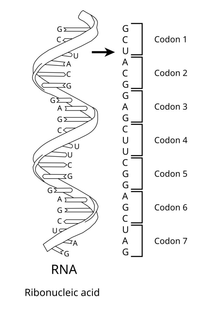
A. Types of Non-Genomic RNA
| Feature | rRNA (Ribosomal) | mRNA (Messenger) | tRNA (Transfer) |
|---|---|---|---|
| Abundance % | $80\%$ (Most abundant). | $2\text{–}5\%$ (Least abundant). | $15\%$. |
| Stability & Size | Most stable; structural ribozyme component. | Shortest lifespan; largest variation in length. | Soluble RNA ($1\text{M HCl}$ soluble); Smallest RNA molecule. |
| Function | Forms ribosome structure and catalyzes peptide bond formation ($23\text{S/28S rRNA}$). | Carries genetic template codon code for protein translation. | Adapter molecule; picks amino acids from cytoplasm and reads mRNA anticodon. |
B. Eukaryotic RNA Polymerases
- RNA Polymerase I: Synthesizes $28\text{S}, 18\text{S}, 5.8\text{S}$ rRNAs.
- RNA Polymerase II: Synthesizes precursor of mRNA called heterogeneous nuclear RNA (hnRNA).
- RNA Polymerase III: Synthesizes tRNA, $5\text{S}$ rRNA, and snRNA (small nuclear RNA).
- Prokaryotes: Single RNA polymerase ($6\text{ polypeptides}$ core enzyme $+$ $\sigma$ factor) transcribes all RNAs.
C. Post-Transcriptional Modifications (Processing of hnRNA $\to$ mRNA)
- 1. Capping: Addition of an unusual nucleotide 7-methyl guanosine triphosphate ($7\text{-mG}_{\text{PPP}}$) at $5’$-end of hnRNA. Protects from nucleases and helps ribosome binding.
- 2. Tailing (Polyadenylation): Addition of 200–300 Adenylate residues (Poly-A tail) at $3’$-end in a template-independent manner.
- 3. Splicing: Removal of non-coding Introns and joining of coding Exons catalyzed by snRNPs (“Snurps” – small nuclear ribonucleoproteins) forming spliceosome.
9. Genetic Code Features & Codon Mathematics
A. Deciphering the Genetic Code
- George Gamow (Physicist): Proposed that genetic code must be a triplet ($4^3 = 64\text{ codons}$) to code for 20 standard amino acids.
- Har Gobind Khorana: Synthesized RNA molecules with defined combinations of homopolymers and copolymers.
- Marshall Nirenberg: Cell-free system for protein synthesis.
- Severo Ochoa Enzyme (Polynucleotide Phosphorylase): Polymerizes RNA without a template.
B. Salient Features of Genetic Code
- 1. Triplet Nature: 61 codons code for amino acids; 3 codons do not code for any amino acid ($\mathbf{UAA\text{ – Ochre}, UAG\text{ – Amber}, UGA\text{ – Opal}}$ = Stop / Nonsense Codons).
- 2. Initiator Codon: AUG has dual functions: codes for Methionine and acts as start codon.
- 3. Unambiguous & Specific: One particular codon codes for only one specific amino acid.
- 4. Degenerate: Some amino acids are coded by more than one codon (e.g., Leucine, Serine, Arginine).
- 5. Comma-less & Non-overlapping: Read continuously without punctuation.
- 6. Universal: From bacteria to humans, $UUU$ codes for Phenylalanine (Exceptions: Mitochondria & Paramecium).
10. tRNA Adapter Model & Ribosome Subunits
A. Structure of tRNA (Adapter Molecule)
- 2D Secondary Structure (Clover Leaf Model – Robert Holley):
• $3’$-Acceptor Arm: Contains $CCA\text{-}3’$ sequence; site for amino acid binding (Aminoacyl attachment).
• Anticodon Loop: Has 7 bases with an anticodon complementary to specific mRNA codon.
• DHU Loop ($D$-loop): Aminoacyl-tRNA synthetase enzyme binding site.
• $T\psi C$ Loop: Ribosome attachment site.
• Variable Loop: Unknown function.
- 3D Tertiary Structure: Compact Inverted L-shaped structure.
B. Ribosome Structure & Translation Sites
Non-membrane bound organelle composed of rRNAs and proteins. Subunits assemble in presence of $\mathbf{Mg^{2+}}$ ions ($0.001\text{ M}$ concentration):
| Ribosome Type | Subunits | rRNA Components & Proteins |
|---|---|---|
| 70S Ribosome (Prokaryotes, Mitochondria, Chloroplast) | Small $30\text{S} +$ Large $50\text{S}$ | • $30\text{S}: 16\text{S rRNA} + 21\text{ proteins}$. • $50\text{S}: 23\text{S rRNA} + 5\text{S rRNA} + 34\text{ proteins}$. • $23\text{S rRNA}$ acts as Ribozyme (Peptidyl transferase). |
| 80S Ribosome (Eukaryotic Cytoplasm) | Small $40\text{S} +$ Large $60\text{S}$ | • $40\text{S}: 18\text{S rRNA} + 33\text{ proteins}$. • $60\text{S}: 28\text{S rRNA} + 5.8\text{S rRNA} + 5\text{S rRNA} + 40\text{ proteins}$. • $28\text{S rRNA}$ acts as Ribozyme. |
C. Translation Binding Sites on Ribosome
- A-site (Aminoacyl site): Accepts incoming charged tRNA.
- P-site (Peptidyl site): Holds tRNA carrying growing polypeptide chain; site where peptide bond is formed.
- E-site (Exit site): Site from where uncharged empty tRNA leaves ribosome.
11. Translation Mechanism & Energy Requirements
A. Overview & Essential Energy Components
Translation is the process of polymerizing amino acids into a polypeptide chain directed by mRNA sequence. Occurs in 4 distinct steps:
| Translation Step | Essential Components & Energy Cost |
|---|---|
| 1. Amino Acid Activation & tRNA Charging | • 1 ATP per amino acid (20 ATP for 20 aa). • Aminoacyl-tRNA Synthetase enzyme & $\text{Mg}^{2+}$ ions. |
| 2. Initiation | • 1 GTP per amino acid. • mRNA, Charged tRNA, $30\text{S} + 50\text{S}$ Ribosome subunits. • Initiation Factors ($IF_1, IF_2, IF_3$), $\text{Mg}^{2+}$ ions. |
| 3. Elongation | • 2 GTP per amino acid added (1 for entry at A-site $+$ 1 for Translocation). • $70\text{S}$ Ribosome unit, Elongation Factors ($EF\text{-Tu}, EF\text{-Ts}, EF\text{-G}$), $\text{Mg}^{2+}$. |
| 4. Termination | • 1 ATP / GTP. • Release Factors ($RF_1, RF_2, RF_3$). |
B. Step 1: Amino Acid Activation & tRNA Charging
Reaction cascade occurring in cytoplasm before ribosome assembly:
$$\text{Amino Acid} + \text{ATP} \xrightarrow[\text{Mg}^{2+}]{\text{Aminoacyl-tRNA Synthetase}} \text{Aminoacyl-AMP-Enzyme Complex} + PP_i$$
$$\text{Complex} + \text{tRNA} \to \mathbf{\text{Aminoacyl-tRNA (Charged tRNA)}} + \text{AMP} + \text{Enzyme}$$
12. Initiation, Elongation & Termination Steps
A. Step 2: Initiation & Shine-Dalgarno Sequence
- $IF_1$ & $IF_3$ bind to $30\text{S}$ small ribosomal subunit.
- Small subunit binds to mRNA at Untranslated Region (UTR) via the Shine-Dalgarno Sequence ($5’\text{-AGGAGG-}3’$ in prokaryotic mRNA complementary to $16\text{S rRNA}$ Anti-Shine Dalgarno sequence $3’\text{-UCCUCC-}5’$).
- Initiating Amino Acid:
• Prokaryotes: N-Formyl Methionine (f-Met).
• Eukaryotes & Archaea: Methionine (Met).
- Initiator tRNA carrying f-Met with anticodon $3’\text{-UAC-}5’$ binds to start codon $5’\text{-AUG-}3’$ directly at P-site (Peptidyl site).
- Large $50\text{S}$ subunit joins forming $70\text{S}$ Initiation Complex ($IF$ factors released).
B. Step 3: Elongation & Translocation
- Next charged tRNA enters empty A-site (Aminoacyl site) driven by Elongation Factors $+$ GTP.
- Peptide bond formed between amino acids at P & A sites by enzyme Peptidyl Transferase ($23\text{S rRNA}$ ribozyme in prokaryotes / $28\text{S rRNA}$ in eukaryotes; non-proteinaceous catalytic RNA).
- Translocation: Ribosome moves 1 codon ahead along mRNA in $5′ \to 3’$ direction. Uncharged tRNA shifts to E-site and leaves; growing peptidyl-tRNA shifts from A-site to P-site.
C. Step 4: Termination
- When a Stop / Nonsense Codon ($UAA, UAG, UGA$) reaches A-site, no tRNA can recognize it.
- Release Factors (RF) bind to stop codon:
• $RF_1$: Recognizes $UAA$ & $UAG$.
• $RF_2$: Recognizes $UAA$ & $UGA$.
• $RF_3$: Helps $RF_1 / RF_2$ release.
- Polypeptide chain is cleaved, tRNA is released, and entire $70\text{S}$ ribosomal machinery dissociates.
13. Regulation of Gene Expression
A. Levels of Gene Regulation in Eukaryotes
Can be regulated at 4 distinct organizational levels:
- Transcriptional Level: Formation of primary transcript / hnRNA (Primary control point).
- Processing Level: Post-transcriptional modifications (Splicing, Capping, Tailing).
- Transport Level: Transport of mature mRNA from nucleus into cytoplasm.
- Translational Level: Polypeptide protein synthesis at ribosome.
B. Levels of Gene Regulation in Prokaryotes
- Predominantly controlled at the Transcriptional Initiation level (e.g., Operon models like Lac Operon).
- Translational control also occurs, but since transcription and translation are coupled in prokaryotic cytoplasm, transcriptional regulation is primary.