1. Biomolecules – Chemical Composition & Metabolites
A. Elemental & Chemical Analysis
- Abundance: $H, C, O, N, S$ are more abundant in living organisms compared to the Earth’s crust, whereas $Na, Mg, Ca, Si$ are higher in Earth’s crust.
- Organic Analysis (Trichloroacetic Acid Test): Living tissue ground with $\text{Cl}_3\text{CCOOH}$ and filtered through cheesecloth/cotton yields two fractions:
- Acid-Soluble Pool (Filtrate): Contains Biomicromolecules (Molecular weight 18 to 800 Daltons). Represents cytoplasm composition without organelles.
- Acid-Insoluble Pool (Retentate): Contains Biomacromolecules (Molecular weight > 10,000 Daltons) such as Proteins, Polysaccharides, Nucleic acids, and organelle membranes.
- Ash Analysis (Inorganic Molecules): Tissue is dried (removes water) and fully burnt to oxidize carbon compounds to gases ($\text{CO}_2, \text{H}_2\text{O}$). Remaining ash contains inorganic elements ($Na, Mg, Ca, K, S, Phosphate, Sulphate$).
B. Secondary Metabolites Table
| Category | Examples |
|---|---|
| Pigments | Carotenoids, Anthocyanins |
| Alkaloids | Morphine, Codeine |
| Terpenoids | Monoterpenes, Diterpenes |
| Essential Oils | Lemon grass oil |
| Toxins | Abrin, Ricin |
| Lectins | Concanavalin A |
| Drugs | Vinblastine, Curcumin |
| Polymer Sub. | Rubber, Gums, Cellulose |
2. Carbohydrates (Saccharides)
Organic polyhydroxy aldehydes or ketones composed of Carbon, Hydrogen, and Oxygen.
A. Monosaccharides ($C_n H_{2n} O_n$)
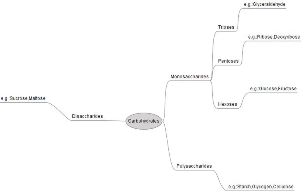
- Simplest carbohydrates that cannot be hydrolyzed further. Contain 3 to 7 Carbon atoms.
- Triose (3C): Glyceraldehyde
- Tetrose (4C): Erythrose
- Pentose (5C): Ribose, Deoxyribose
- Hexose (6C): Glucose, Fructose, Galactose
- Heptose (7C): Sedoheptulose
- Optical Activity: Dextrorotatory ($+$ / rotates light right e.g., Glucose) & Levorotatory ($-$ / rotates light left e.g., Fructose).
- Structural Rings: Glucose forms a 6-membered Pyranose ring; Ribose & Fructose form a 5-membered Furanose ring.
- Derived Sugars: Deoxy-sugars (Deoxyribose – removal of oxygen from 2nd Carbon), Amino-sugars (Glucosamine), Sugar Acids (Ascorbic acid, Gluronic acid), Sugar Alcohols (Mannitol in brown algae).
B. Oligosaccharides & Sugar Tests
- Formed by condensation of 2 to 9 monosaccharide units joined by Glycosidic Bonds (with loss of $\text{H}_2\text{O}$).
- Disaccharide Examples:
- Maltose: $\alpha\text{-Glucose} + \alpha\text{-Glucose}$ ($\alpha 1,4\text{-glycosidic bond}$).
- Sucrose: $\text{Glucose} + \text{Fructose}$ ($\alpha 1, \beta 2\text{-glycosidic bond}$). Non-reducing sugar because free aldehyde/ketone group is absent.
- Lactose: $\text{Glucose} + \text{Galactose}$ ($\beta 1,4\text{-glycosidic bond}$). Milk sugar.
- Trisaccharide Example: Raffinose ($\text{Glucose} + \text{Fructose} + \text{Galactose}$).
- Benedict’s / Fehling’s Test: All monosaccharides and maltose/lactose reduce $\text{Cu}^{2+}$ (blue) to $\text{Cu}^+$ (brick red precipitate) due to free functional groups $\to$ Reducing Sugars.
3. Polysaccharides & Amino Acids
A. Polysaccharides (Complex Carbohydrates)
Acid-insoluble macromolecular polymers. Chemically and osmotically inactive, making them ideal for storage.
- Homopolysaccharides: Made of single type of monomer.
- Starch: Plant reserve food; Polymer of $\alpha$-glucose. Composed of Amylose (unbranched, helical, $\alpha 1,4$ bonds; gives Blue-Black color with $I_2$) and Amylopectin (branched, $\alpha 1,4$ and $\alpha 1,6$ bonds; gives Reddish-Violet color with $I_2$).
- Glycogen: Animal reserve food stored in liver and muscle; highly branched polymer of $\alpha$-glucose. Gives Brick-Red color with $I_2$.
- Cellulose: Unbranched homopolymer of $\beta$-glucose ($\beta 1,4$ bonds); structural component of plant cell walls and cotton fibers (90% cellulose). Does not hold $I_2$ molecules.
- Chitin: Homopolymer of $N$-Acetylglucosamine (NAG) linked by $\beta 1,4$ bonds. Found in fungal cell walls and arthropod exoskeletons.
- Inulin: Polymer of fructose; reserve food in Sunflower family. Used in clinical testing for Kidney Glomerular Filtration Rate (GFR) as it is neither metabolized nor reabsorbed.
- Heteropolysaccharides: Made of different monomeric units (e.g., Peptidoglycan, Hyaluronic acid, Agar-Agar).
B. Amino Acids Structure & Types
Substituted methanes containing an amino group ($-\text{NH}_2$), acidic carboxyl group ($-\text{COOH}$), hydrogen, and variable functional group ($-\text{R}$) attached to $\alpha$-carbon.
- Chemical Classification:
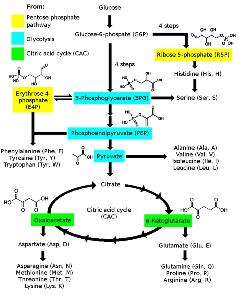
- Neutral: $\text{NH}_2 = \text{COOH}$ (e.g., Glycine, Alanine, Valine, Leucine, Isoleucine).
- Acidic: $\text{COOH} > \text{NH}_2$ (e.g., Glutamic acid, Aspartic acid).
- Basic: $\text{NH}_2 > \text{COOH}$ (e.g., Lysine, Arginine, Histidine).
4. Amino Acid Properties & Protein Structure
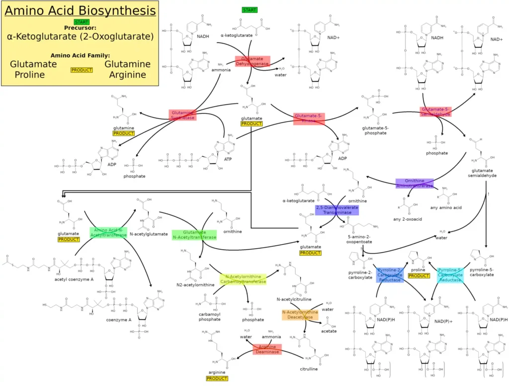
A. Special Amino Acids & Zwitterion
- Sulphur-containing: Cysteine, Methionine.
- Aromatic: Phenylalanine, Tryptophan, Tyrosine.
- Heterocyclic: Proline, Hydroxyproline, Histidine.
- Zwitterion Property: At physiological pH (7.4), amino acids carry both positive and negative charges simultaneously ($\text{H}_3\text{N}^+ – \text{CH(R)} – \text{COO}^-$).
- Basic pH $\to$ Exists as Anion (Acidic Protein e.g., Plasma).
- Acidic pH $\to$ Exists as Cation (Basic Protein e.g., Histones).
- Nutritional Classification: 20 Standard Amino Acids.
- Essential (From Diet): Phenylalanine, Valine, Tryptophan, Threonine, Isoleucine, Methionine, Histidine, Arginine, Lysine, Leucine (Mnemonic: PVT TIM HALL).
- Non-Essential: Synthesized by body (e.g., Glycine, Alanine).
B. Protein Structural Levels
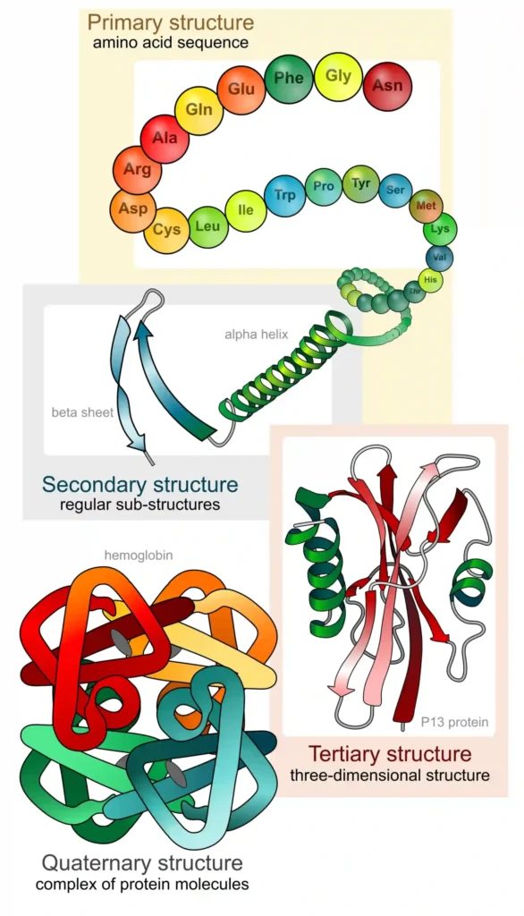
Proteins are heteropolymers of amino acids linked by Peptide Bonds ($-\text{CO}-\text{NH}-$ formed by dehydration). Number of peptide bonds = $n – 1$.
- 1. Primary Structure: Linear sequence of amino acids giving positional information. Has $N$-terminal (first amino acid) and $C$-terminal (last amino acid). Positional specific but non-functional.
- 2. Secondary Structure: Local folding of primary chain stabilized by Hydrogen bonds.
- $\alpha$-Helix: Right-handed helix with intramolecular H-bonds (e.g., Keratin in hair).
- $\beta$-Pleated Sheet: Left/Right handed with intermolecular H-bonds (e.g., Silk Fibroin).
- Triple Helix: Three polypeptide chains coiled together (e.g., Collagen – described by G.N. Ramachandran).
5. Advanced Protein Structure & Lipids
A. Tertiary & Quaternary Protein Structure
- 3° Structure (Tertiary): Long polypeptide chain folds upon itself like a hollow woollen ball forming a 3D structure.
- Stabilized by Peptide, Hydrogen, Ionic, Disulphide, and Hydrophobic bonds.
- Creates Active Sites essential for biological/enzymatic activity (e.g., Myoglobin, Globular proteins).
- 4° Structure (Quaternary): Architecture of proteins composed of more than one polypeptide subunit (e.g., Adult Haemoglobin with $2\alpha$ and $2\beta$ chains).
• Most abundant protein in Animal World: Collagen
• Most abundant protein in Whole Biosphere: RuBisCO (Ribulose bisphosphate carboxylase-oxygenase).
B. Lipids Classification
Esters of fatty acids and alcohol; water insoluble but soluble in non-polar organic solvents (benzene, ether).
- 1. Simple Lipids: Esters of fatty acid + alcohol (Glycerol).
- Triglycerides: $1\text{ Glycerol} + 3\text{ Fatty Acids}$ linked by Ester bonds.
- Saturated Fatty Acids: No double bonds; higher melting point; solid at room temp (e.g., Palmitic acid – 16C, Stearic acid – 18C).
- Unsaturated Fatty Acids: Contains 1 or more double bonds ($= $); low melting point; liquid at room temp. MUFA (Oleic acid – 18C, 1 double bond) & PUFA (Linoleic acid – 18C 2=, Linolenic acid – 18C 3=, Arachidonic acid – 20C 4=).
6. Lipids (Contd.) & Nucleic Acids
A. Conjugated & Derived Lipids
- Conjugated Lipids: Fatty acid + Alcohol + Non-lipid group.
- Phospholipids: Glycerol + 2 Fatty Acids + Phosphate group linked to Choline. Example: Lecithin (found in cell membranes and alveolar lining of lungs to reduce surface tension). Amphipathic nature (Hydrophilic polar head + Hydrophobic non-polar tails).
- Glycolipids: Fatty acid + Sphingosine alcohol + Sugar (e.g., Cerebrosides).
- Lipoproteins: Lipid + Protein.
- Derived Lipids: Hydrolysis products of simple/complex lipids possessing lipid-like properties (e.g., Sterols / Cholesterol synthesized in liver; Prostaglandins derived from Arachidonic acid).
B. Nucleic Acids (DNA & RNA)
Largest biomolecules; acid-insoluble polymers of Nucleotides.
- Components of Nucleotide:
- Pentose Sugar: Ribose (in RNA) or 2′-Deoxyribose (in DNA).
- Nitrogenous Base: Purines (2 rings: Adenine, Guanine – nitrogen at 1, 3, 7, 9 positions) & Pyrimidines (1 ring: Cytosine, Thymine in DNA, Uracil in RNA – nitrogen at 1, 3 positions).
- Phosphate Group: Linked to sugar via Phosphoester bond.
- Nucleoside vs Nucleotide:
- $\text{Sugar} + \text{N-Base} = \text{Nucleoside}$ (e.g., Adenosine, Cytidine, Guanosine, Uridine). Linked via $N$-Glycosidic bond.
- $\text{Nucleoside} + \text{Phosphate} = \text{Nucleotide}$ (e.g., Adrenylic acid, Cytidylic acid).
- DNA Base Pairing: Double-stranded helix connected by Hydrogen bonds ($A = T$ with 2 H-bonds, $G \equiv C$ with 3 H-bonds).
7. Enzymes & Catalytic Action
Biological catalysts, mostly proteins with 3° structure forming active site pockets (Exceptions: Ribozyme, Ribonuclease P are nucleic acid enzymes).
A. Enzymatic Mechanism & Kinetics
- Enzymes increase reaction rates dramatically by lowering Activation Energy required to reach the transition state.
- Rate of Reaction ($\text{Rate} = \frac{dP}{dT}$): Increases 2-fold for every 10°C rise in temperature until denaturation point.Example: $\text{CO}_2 + \text{H}_2\text{O} \xrightleftharpoons{\text{Carbonic Anhydrase}} \text{H}_2\text{CO}_3$ produces 600,000 molecules/sec with enzyme vs 200 molecules/hour without enzyme ($10^7$ times faster).
- Thermostable Enzymes: Isolated from thermophilic organisms (e.g., Taq Polymerase from Thermus aquaticus retains activity at >90°C).
B. Factors Affecting Enzyme Activity
- 1. Temperature & pH: Enzymes show peak activity at optimum temperature (25–40°C) and optimum pH. Low temperature temporarily inactives enzyme; high temperature denatures protein structure permanently.
- 2. Substrate Concentration ($[S]$): Reaction velocity forms a Rectangular Hyperbola.
- Michaelis-Menten Constant ($K_m$): Substrate concentration at which reaction velocity reaches half of maximum velocity ($\frac{V_{max}}{2}$).
- Low $K_m \to$ High affinity of enzyme for substrate; High $K_m \to$ Low affinity ($K_m \propto \frac{1}{\text{Affinity}}$).