Quick Summary: Cell Theory, Discoveries, Prokaryotic & Eukaryotic Cell structures, Gram Staining, Plasma Membrane models, Endomembrane System (ER, Golgi, Lysosomes, Vacuoles), Mitochondria, Plastids, Ribosomes, Cytoskeleton, Cilia/Flagella (9+2 & 9+0 array), Centrosome, and Nucleus structure.
1. Cell – Introduction & Key Discoveries
A. Major Biological Discoveries
- G.N. Ramachandran: Triple helical structure of collagen (inspired by Linus Pauling).
- Matthias Schleiden (1838): German botanist, concluded plants are composed of cells.
- Theodore Schwann (1839): German zoologist, observed plasma membrane and noted cell wall is unique to plant cells.
- Rudolf Virchow (1855): Stated “Omnis cellula-e cellula” (All cells arise from pre-existing cells). Extended Cell Theory.
- Singer & Nicolson (1972): Proposed Fluid Mosaic Model of plasma membrane.
- Camillo Golgi (1898): Discovered Golgi body.
- George Palade (1953): Discovered Ribosomes.
- Robert Brown (1831): Discovered Nucleus.
- Flemming: Named stained chromatin in nucleus.
- J.E. Purkinje: Coined the term Protoplasm.
B. Cell Theory & Cell Types
Cell Theory: Proposed by Schleiden & Schwann, modified by Virchow. Exceptions: Viruses, Viroids, Prions.
- Smallest Cell: Mycoplasma (0.3 µm)
- Bacteria Size: 3 – 5 µm
- RBC Size: 7 µm (Biconcave & Round)
- Longest Cell: Neuron
- Largest Cell: Ostrich Egg
2. Prokaryotic Cells
Includes Bacteria, Blue-Green Algae (BGA), Mycoplasma. Multiply rapidly, smaller in size.
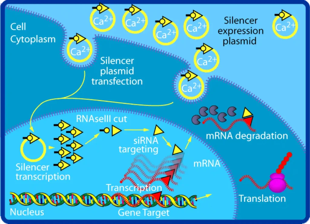
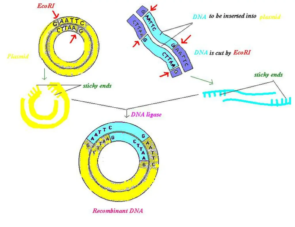
SPECIAL NOTE – PLASMID: Extra-chromosomal, small, circular, double-stranded DNA that gives unique phenotypic characters to bacteria (e.g., resistance to antibiotics).
Cell Envelope Structures
- Glycocalyx (Outermost): Loose sheath = Slime layer; Thick and tough = Capsule. Protects cell from host immune system.
- Cell Wall: Made of Peptidoglycan (murein). Provides shape and structural support, prevents cell from bursting or collapsing.
- Plasma Membrane: Selectively permeable, lipid bilayer structure.
- Mesosome: Infoldings of plasma membrane. Helps in cell wall formation, DNA replication, distribution to daughter cells, respiration, and increasing enzymatic surface area.
- Polysome / Polyribosome: Multiple 70S ribosomes attached to a single mRNA strand for translation.
- Chromatophore: Pigment-containing membrane extensions in photosynthetic prokaryotes (e.g., BGA, Purple/Green Sulphur bacteria).
3. Gram Staining & Eukaryotic Comparison
A. Gram Staining Technique (Christian Gram)
Differentiates bacteria based on cell wall composition using Crystal Violet stain, Iodine, Alcohol wash, and Safranin counterstain.
| Feature | Gram Positive (+ve) | Gram Negative (-ve) |
|---|---|---|
| Stain Retention | Retains Crystal Violet (Violet/Blue) | Takes Safranin Counterstain (Pink) |
| Peptidoglycan Layer | Thick single layer | Thin layer + Outer Lipopolysaccharide membrane |
| Teichoic Acid | Present | Absent |
| Examples | Streptococcus, Staphylococcus | E. coli, Rhizobium |
B. Prokaryote vs. Eukaryote Differences
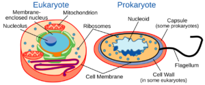
- Prokaryotes: Nuclear envelope absent; G-C rich circular DNA without histones; 70S ribosomes; membrane-bound organelles absent.
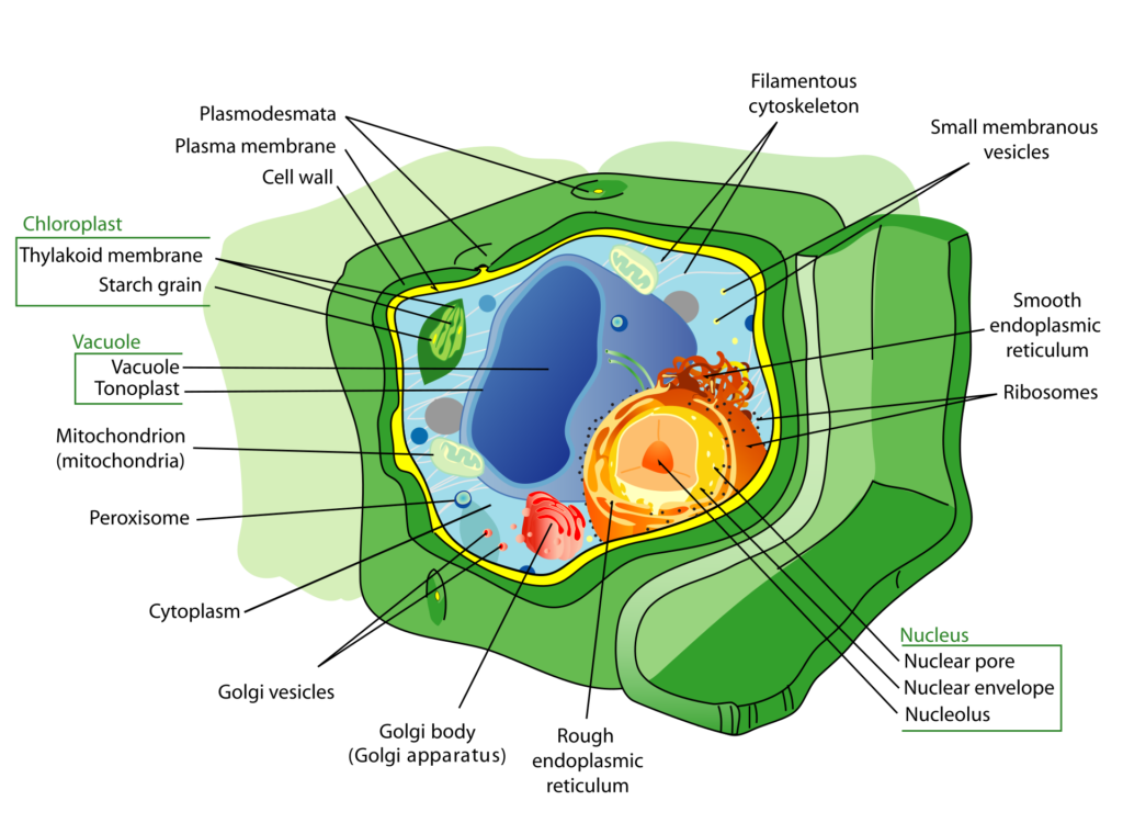
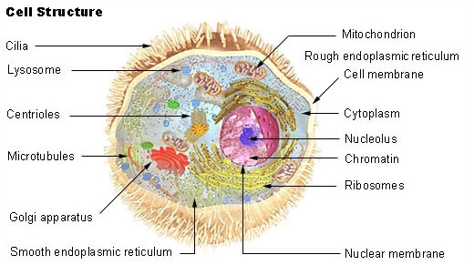
- Eukaryotes: Distinct nuclear envelope present; Linear DNA with histones (A-T rich); 80S ribosomes (cytoplasm); Membrane-bound organelle system present.
C. Cell Wall Variations
- Algae: Made of Cellulose, Galactans, Mannans, and Minerals ($CaCO_3$).
- Plants: Cellulose, Hemicellulose, Pectins, and Proteins.
- Middle Lamella: Made of Calcium/Magnesium pectate; cements neighboring cells together.
4. Plasma Membrane & Endomembrane System
A. Plasma Membrane Structure & Transport
- Fluid Mosaic Model (Singer & Nicolson): Quasi-fluid nature of lipids enables lateral movement of proteins within the overall bilayer.
- Lipid Composition: Phosphoglycerides with polar head facing outwards (hydrophilic) and non-polar saturated hydrocarbon tail facing inwards (hydrophobic). Human RBC membrane has 52% protein and 40% lipid.
- Membrane Proteins: Integral (Intrinsic / Transmembrane) and Peripheral (Extrinsic – easy to extract).
- Transport Types:
- Passive Transport: Along concentration gradient without energy expenditure (e.g., Simple Diffusion & Osmosis).
- Facilitated Diffusion: Neutral/polar molecules cross via membrane carrier proteins without energy requirement.
- Active Transport: Against concentration gradient using ATP (e.g., $Na^+ / K^+$ Pump).
- Bulk Transport: Endocytosis (Phagocytosis = cell eating, Pinocytosis = cell drinking) & Exocytosis (Cell vomiting).
B. Endoplasmic Reticulum (ER)
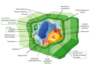
Network of tiny tubular structures dividing intracellular space into Luminal (inside ER) and Extra-luminal (cytoplasm) compartments.
- Rough ER (RER): Bears 80S ribosomes on outer surface; active in protein synthesis and secretion (e.g., Pancreatic cells, Brain cells). Attached via Ribophorin I & II.
- Smooth ER (SER): Lacks ribosomes; major site for synthesis of lipids, steroidal hormones (testosterone), and glycogen metabolism/detoxification in liver.
5. Golgi Apparatus, Lysosome, Vacuole & Mitochondria
A. Golgi Body (Camillo Golgi, 1898)
- Consists of flattened disc-shaped sacs called Cisternae (0.5 – 1.0 µm diameter) stacked parallelly. In plant cells, referred to as Dictyosomes.
- Distinct Faces: Cis face (Convex / Forming face toward nucleus) and Trans face (Concave / Maturing face toward plasma membrane).
- Primary Functions: Modification of proteins (Glycosylation) and lipids (Glycolipidation); packaging and sorting materials for secretion; formation of Acrosome in sperm.
B. Lysosomes (“Suicidal Bags”)
- Single membrane-bound vesicular structures formed by packaging in Golgi apparatus.
- Rich in hydrolytic enzymes (lipases, proteases, carbohydrates) active at acidic pH (maintained by active proton pumping).
- Exhibits Polymorphism: Primary lysosomes, Secondary (Heterophagosome), Residual bodies, and Autophagic vacuoles.
C. Vacuoles & Mitochondria
- Vacuole: Bound by single membrane Tonoplast. Occupies up to 90% volume of plant cells. Tonoplast facilitates transport against concentration gradient.
- Contractile Vacuole: Osmoregulation/excretion in Amoeba.
- Food Vacuole: Engulfs food particles in Protista.
- Mitochondria (“Powerhouse of Cell”): Double-membrane organelle (Sausage/Cylindrical shaped, 0.2 – 1.0 µm diameter).
- Outer membrane is smooth; Inner membrane forms infoldings called Cristae to increase surface area.
- Matrix contains single circular dsDNA, 70S ribosomes, RNA, and Krebs cycle enzymes. Stained with Janus Green.
- Divides by binary fission. Semi-autonomous organelle.
6. Plastids, Ribosomes & Cytoskeleton
A. Plastids Classification
| Plastid Type | Color / Pigments | Function / Storage |
|---|---|---|
| Leucoplast | Colourless | • Amyloplast: Stores Starch (Potato, Rice) • Elaioplast: Stores Fats/Oils (Castor) • Aleuroplast: Stores Proteins (Maize) |
| Chromoplast | Yellow, Orange, Red (Carotenoids, Xanthophylls) | Gives characteristic color to fruits, flowers (Chilli, Tomato) |
| Chloroplast | Green (Chlorophyll & Carotenoids) | Site of Photosynthesis. Contains flattened thylakoid stacks (Grana) and Stroma. |
B. Ribosomes & Cytoskeleton
- Ribosomes (Protein Factories): Non-membrane bound granules made of rRNA and proteins.
- 70S Type: 50S + 30S subunits (In Prokaryotes, Mitochondria, Chloroplasts). Attached via $Mg^{2+}$ ions.
- 80S Type: 60S + 40S subunits (In Eukaryotic cytoplasm).
- ‘S’ = Svedberg’s Unit (Sedimentation Coefficient).
- Cytoskeleton: Elaborate network of filamentous proteinaceous structures in cytoplasm. Composed of Microtubules, Microfilaments, and Intermediate Filaments. Provides mechanical support, motility, and maintenance of cell shape.
7. Cilia, Flagella, Centrosome & Nucleus
A. Cilia & Flagella Arrangement
- Hair-like outgrowth of plasma membrane. Core is called Axoneme.
- (9 + 2) Array Structure: 9 peripheral doublet microtubules + 2 central singlet microtubules connected by radial spokes and interdoublet bridges (Nexin protein).
- Both cilia and flagella emerge from centriole-like structure called Basal body.
B. Centrosome & Centrioles
- Membraneless organelle containing 2 cylindrical structures called Centrioles lying perpendicular to each other.
- (9 + 0) Cartwheel Arrangement: Made of 9 evenly spaced peripheral triplets of Tubulin protein (Total 27 microtubules per centriole). Central region is proteinaceous Hub connected to spokes.
C. Nucleus Structure & Chromatin
- Double membrane-bound organelle with nuclear pores forming passage for RNA and proteins. Perinuclear space is 10 – 50 nm.
- Nucleolus: Non-membrane bound spherical structure in nucleoplasm; active site for ribosomal RNA (rRNA) synthesis.
- Chromatin Types:
- Euchromatin: Loosely packed, transcriptionally active, stains light.
- Heterochromatin: Tightly packed, transcriptionally inactive, stains dark.